

购物车
 您的购物车当前为空

 很棒
很棒Ruxolitinib (INCB018424) 是一种 JAK1/2 抑制剂 (IC50=3.3/2.8 nM),具有有效性和选择性。Ruxolitinib 具有抗肿瘤活性,可以诱导细胞自噬和凋亡。
很棒纯度: 99.99%
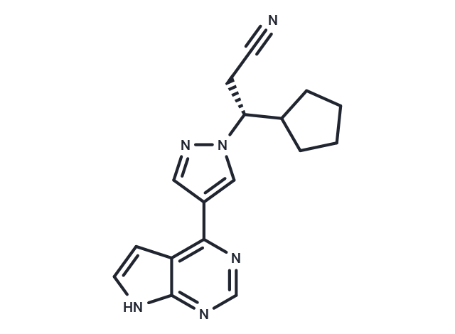
别名 鲁索替尼, 鲁索利替尼, 芦可替尼, INCB018424, (R)-Ruxolitinib
Ruxolitinib (INCB018424) 是一种 JAK1/2 抑制剂 (IC50=3.3/2.8 nM),具有有效性和选择性。Ruxolitinib 具有抗肿瘤活性,可以诱导细胞自噬和凋亡。
| 规格 | 价格 | 库存 | 数量 |
|---|---|---|---|
| 1 mg | ¥ 233 | 现货 | |
| 5 mg | ¥ 536 | 现货 | |
| 10 mg | ¥ 828 | 现货 | |
| 25 mg | ¥ 1,530 | 现货 | |
| 50 mg | ¥ 2,320 | 现货 | |
| 100 mg | ¥ 3,730 | 现货 | |
| 200 mg | ¥ 4,990 | 现货 | |
| 500 mg | ¥ 7,690 | 现货 | |
| 1 mL x 10 mM (in DMSO) | ¥ 611 | 现货 |
TargetMol的所有产品仅用作科学研究或药证申报,不能被用于人体,我们不向个人提供产品和服务。请您遵守承诺用途,不得违反法律法规规定用于任何其他用途。
凭借在化合物合成方面的丰富经验,我们可以根据您的研究需求为该产品提供快速定制合成服务。
| 产品描述 | Ruxolitinib (INCB018424) is a JAK1/2 inhibitor (IC50=3.3/2.8 nM) that is potent and selective. Rixolitinib exhibits antitumor activity and induces autophagy and apoptosis. |
| 靶点活性 | TYK2:19 nM (cell free), JAK1:3.3 nM (cell free), JAK2:2.8 nM (cell free), JAK3:428 nM |
| 体外活性 | 方法:Ba/F3-EpoR-JAK2V617F 细胞用 Ruxolitinib (0-10 μM) 处理 48 h,使用 Cell-Titer Glo 检测的细胞活力。 |
| 体内活性 | 方法:为检测体内抗肿瘤活性,将 Ruxolitinib (3-30 mg/kg,5% dimethyl acetamide, 0.5% methocellulose) 灌胃给药给携带肿瘤 Ba/F3-JAK2V617F 的 BALB/c 小鼠,每天两次,持续三周。 |
| 激酶实验 | The kinase domains of human JAK1 (837-1142), JAK2 (828-1132), JAK3 (781-1124), and Tyk2 (873-1187) were cloned by PCR with N-terminal epitope tags. Recombinant proteins were expressed using Sf21 cells and baculovirus vectors and purified with affinity chromatography. JAK kinase assays used a homogeneous time-resolved fluorescence assay with the peptide substrate (-EQEDEPEGDYFEWLE). Each enzyme reaction was carried out with test compound or control, JAK enzyme, 500nM peptide, adenosine triphosphate (ATP; 1mM), and 2.0% dimethyl sulfoxide (DMSO) for 1 hour. The 50% inhibitory concentration (IC50) was calculated as the compound concentration required for inhibition of 50% of the fluorescent signal. Biochemical assays for CHK2 and c-MET enzymes were performed using standard conditions (Michaelis constant [Km] ATP) with recombinantly expressed catalytic domains from each protein and synthetic peptide substrates. An additional panel of kinase assays (Abl, Akt1, AurA, AurB, CDC2, CDK2, CDK4, CHK2, c-kit, c-Met, EGFR, EphB4, ERK1, ERK2, FLT-1, HER2, IGF1R, IKKα, IKKβ, JAK2, JAK3, JNK1, Lck, MEK1, p38α, p70S6K, PKA, PKCα, Src, and ZAP70) was performed using standard conditions using 200nM INCB018424. Significant inhibition was defined as more than or equal to 30% (average of duplicate assays) compared with control values [1]. |
| 细胞实验 | Cells were seeded at 2000/well of white bottom 96-well plates, treated with compounds from DMSO stocks (0.2% final DMSO concentration), and incubated for 48 hours at 37°C with 5% CO2. Viability was measured by cellular ATP determination using the Cell-Titer Glo luciferase reagent or viable cell counting. Values were transformed to percent inhibition relative to vehicle control, and IC50 curves were fitted according to nonlinear regression analysis of the data using PRISM GraphPad [1]. |
| 动物实验 | All of the procedures were conducted in accordance with the US Public Health Service Policy on Humane Care and Use of Laboratory Animals. Mice were fed standard rodent chow and provided with water ad libitum. Ba/F3-JAK2V617F cells (10^5 per mouse) were inoculated intravenously into 6- to 8-week-old female BALB/c mice. Survival was monitored daily, and moribund mice were humanely killed and considered deceased at time of death. Treatment with vehicle (5% dimethylacetamide, 0.5% methocellulose) or INCB018424 began within 24 hours of cell inoculation, twice daily by oral gavage. Hematologic parameters were measured using a Bayer Advia120 analyzed, and statistical significance was determined using Dunnett testing [1]. |
| 别名 | 鲁索替尼, 鲁索利替尼, 芦可替尼, INCB018424, (R)-Ruxolitinib |
| 分子量 | 306.36 |
| 分子式 | C17H18N6 |
| CAS No. | 941678-49-5 |
| Smiles | N#CC[C@H](C1CCCC1)n1cc(cn1)-c1ncnc2[nH]ccc12 |
| 密度 | 1.40g/cm3 |
| 存储 | Store at low temperature Powder: -20°C for 3 years | In solvent: -80°C for 1 year Shipping with blue ice/Shipping at ambient temperature. 实际储存温度请以COA为准 | |||||||||||||||||||||||||||||||||||
| 溶解度信息 | H2O: < 1 mg/mL (insoluble or slightly soluble) DMSO: 242 mg/mL (789.92 mM), Sonication is recommended. Ethanol: < 1 mg/mL (insoluble or slightly soluble) | |||||||||||||||||||||||||||||||||||
| 体内实验配方 | 10% DMSO+40% PEG300+5% Tween 80+45% Saline: 5.7 mg/mL (18.61 mM), Solution. 请按顺序添加溶剂,在添加下一种溶剂之前,尽可能使溶液澄清。如有必要,可通过加热、超声、涡旋处理进行溶解。工作液建议现配现用。以上配方仅供参考,体内配方并不是绝对的,请根据不同情况进行调整。 | |||||||||||||||||||||||||||||||||||
溶液配制表 | ||||||||||||||||||||||||||||||||||||
DMSO
该溶液配制表仅适用于固体产品。对于液体产品,请根据标明的浓度或密度计算稀释方案。 | ||||||||||||||||||||||||||||||||||||
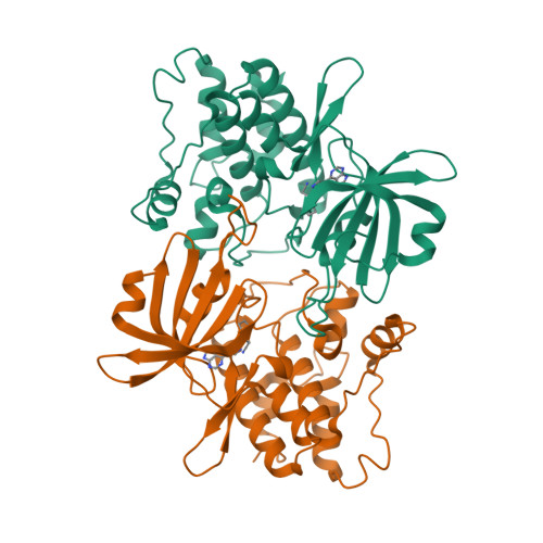
C-Src in complex with Ruxolitinib
对于不同动物的给药剂量换算,您也可以参考 更多
 嗨!有任何问题?点我咨询
版权所有©2015-2026 TargetMol Chemicals Inc.保留所有权利.
沪ICP备20019793号-4 | 沪公网安备 31010602006700号 | 沪(静)应急管危经许[2024]203441