

购物车
 您的购物车当前为空


 还可以
还可以Anti-PARK7/DJ-1 Polyclonal Antibody 是一种 Rabbit 抗体,靶向 PARK7/DJ-1。Anti-PARK7/DJ-1 Polyclonal Antibody 可用于 FCM,IF,IHC-Fr,IHC-P,WB。
还可以别名 parkinson protein 7, HEL-S-67p, DJ-1, DJ1
Anti-PARK7/DJ-1 Polyclonal Antibody 是一种 Rabbit 抗体,靶向 PARK7/DJ-1。Anti-PARK7/DJ-1 Polyclonal Antibody 可用于 FCM,IF,IHC-Fr,IHC-P,WB。
| 规格 | 价格 | 库存 | 数量 |
|---|---|---|---|
| 50 μL | ¥ 1,160 | 5日内发货 | |
| 100 μL | ¥ 1,960 | 5日内发货 | |
| 200 μL | ¥ 2,790 | 5日内发货 |
TargetMol的所有产品仅用作科学研究或药证申报,不能被用于人体,我们不向个人提供产品和服务。请您遵守承诺用途,不得违反法律法规规定用于任何其他用途。
| 产品描述 | Anti-PARK7/DJ-1 Polyclonal Antibody is a Rabbit antibody targeting PARK7/DJ-1. Anti-PARK7/DJ-1 Polyclonal Antibody can be used in FCM,IF,IHC-Fr,IHC-P,WB. |
| 别名 | parkinson protein 7, HEL-S-67p, DJ-1, DJ1 |
| Ig Type | IgG |
| 反应种属 | Human,Mouse,Rat (predicted:Pig,Cow,Horse) |
| 验证活性 | 1. Blank control: 293T cells (blue). Primary Antibody: Rabbit Anti-PARK7/CAP1 antibody (TMAB-01332), Dilution: 0.2 μg in 100 μL 1X PBS containing 0.5% BSA; Isotype Control Antibody: Rabbit IgG (orange),used under the same conditions. Secondary Antibody: Goat anti-rabbit IgG-Pe (white blue), Dilution: 1:200 in 1 X PBS containing 0.5% BSA. Protocol Primary antibody (TMAB-01332, 0.2 μg/1x10^6 cells) were incubated for 30 min on the ice, followed by 1 X PBS containing 0.5% BSA + 10% goat serum (15 min) to block non-specific protein-protein interactions. Then the Goat Anti-rabbit IgG/PE antibody was added into the blocking buffer mentioned above to react with the primary antibody at 1/200 dilution for 30 min on ice. 2. Tissue/cell: rat kidney tissue; 4% Paraformaldehyde-fixed and paraffin-embedded; Antigen retrieval: citrate buffer (0.01M, pH6.0), Boiling bathing for 15 min; Block endogenous peroxidase by 3% Hydrogen peroxide for 30 min; Blocking buffer (normal goat serum) at 37°C for 20 min; Incubation: Anti-CAP1/PARK7 Polyclonal Antibody, Unconjugated (TMAB-01332) 1:200, overnight at 4°C, followed by conjugation to the secondary antibody and DAb staining. 3. Tissue/cell: rat kidney tissue; 4% Paraformaldehyde-fixed and paraffin-embedded; Antigen retrieval: citrate buffer (0.01M, pH6.0), Boiling bathing for 15 min; Block endogenous peroxidase by 3% Hydrogen peroxide for 30 min; Blocking buffer (normal goat serum) at 37°C for 20 min; Incubation: Anti-CAP1/PARK7 Polyclonal Antibody, Unconjugated (TMAB-01332) 1:200, overnight at 4°C, followed by conjugation to the secondary antibody and DAb staining. 4. The blue histogram is unstained cells (Hepg2 cells) concentration 1:50 The Wathet Blue histogram is cells stained with secondary antibody alone. The Orange histogram is cells stained with rabbit IgG isotype control antibody plus secondary antibody. The green histogram is cells stained with Rabbit Anti-PARK7/CAP1 antibody (TMAB-01332) plus secondary antibody. 5. Sample: Lane 1: Testis (Mouse) Lysate at 40 μg Lane 2: Liver (Mouse) Lysate at 40 μg Lane 3: Cerebrum (Rat) Lysate at 40 μg Lane 4: Thyroid gland Rat) Lysate at 40 μg Lane 5: Kidney (Rat) Lysate at 40 μg Lane 6: Liver (Rat) Lysate at 40 μg Lane 7: Hela (Human) Cell Lysate at 30 μg Lane 8: U937 (Human) Cell Lysate at 30 μg Lane 9: K562 (Human) Cell Lysate at 30 μg Lane 10: HL60 (Human) Cell Lysate at 30 μg Primary: Anti-PARK7/DJ1 (TMAB-01332) at 1/1000 dilution Secondary: IRDye800CW Goat Anti-Rabbit IgG at 1/20000 dilution Predicted band size: 22 kDa Observed band size: 22 kDa 6. Paraformaldehyde-fixed, paraffin embedded (human colon carcinoma); Antigen retrieval by boiling in sodium citrate buffer (pH6.0) for 15 min; Block endogenous peroxidase by 3% hydrogen peroxide for 20 min; Blocking buffer (normal goat serum) at 37°C for 30 min; Antibody incubation with (PARK7) Polyclonal Antibody, Unconjugated (TMAB-01332) at 1:200 overnight at 4°C, followed by operating according to SP Kit (Rabbit) instructionsand DAB staining. 7. Paraformaldehyde-fixed, paraffin embedded (rat brain); Antigen retrieval by boiling in sodium citrate buffer (pH6.0) for 15 min; Block endogenous peroxidase by 3% hydrogen peroxide for 20 min; Blocking buffer (normal goat serum) at 37°C for 30 min; Antibody incubation with (PARK7) Polyclonal Antibody, Unconjugated (TMAB-01332) at 1:200 overnight at 4°C, followed by operating according to SP Kit (Rabbit) instructionsand DAB staining. 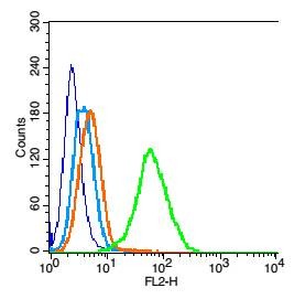 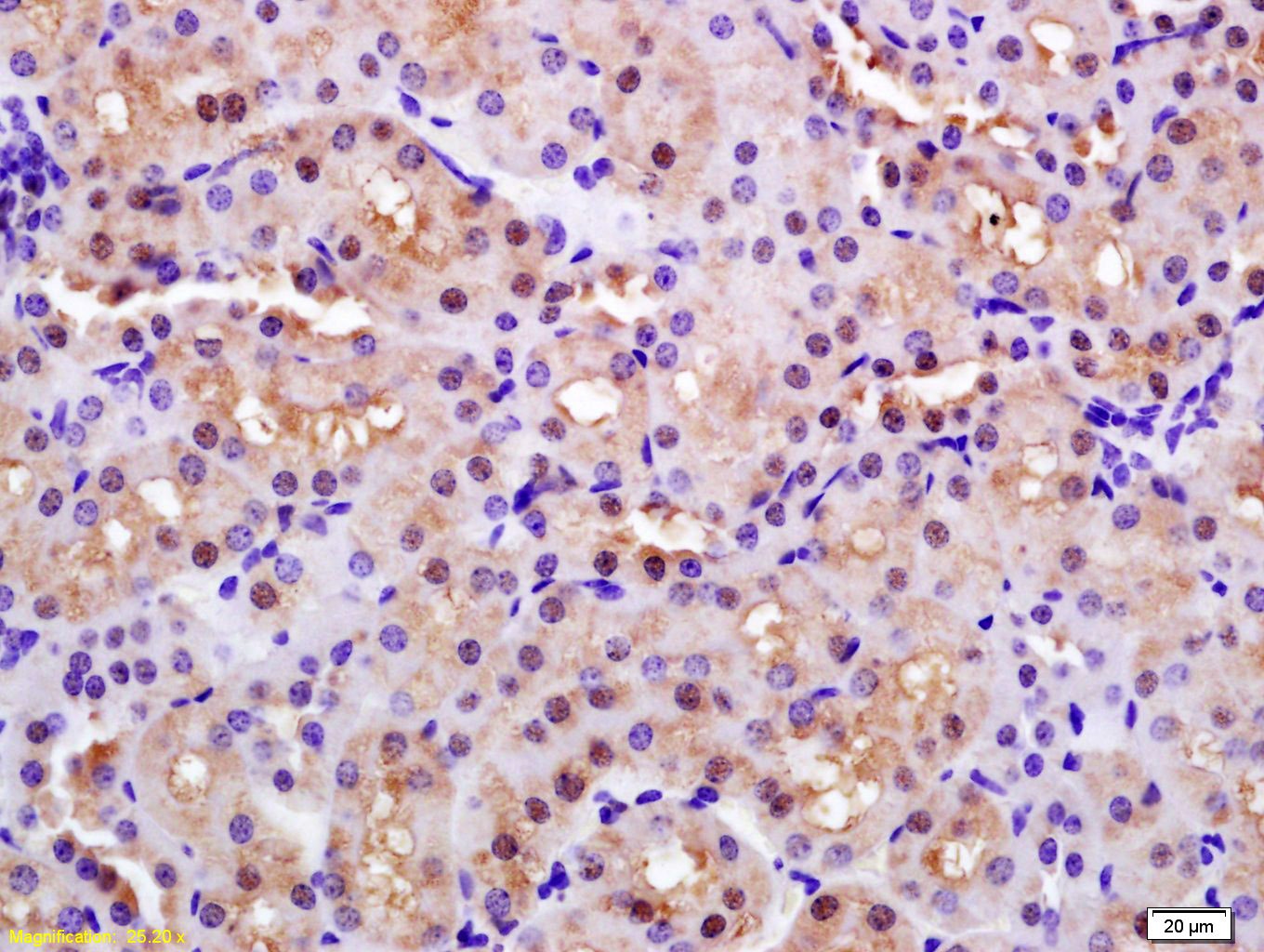 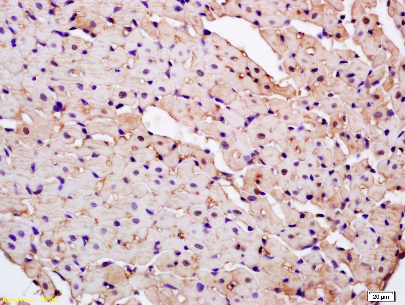 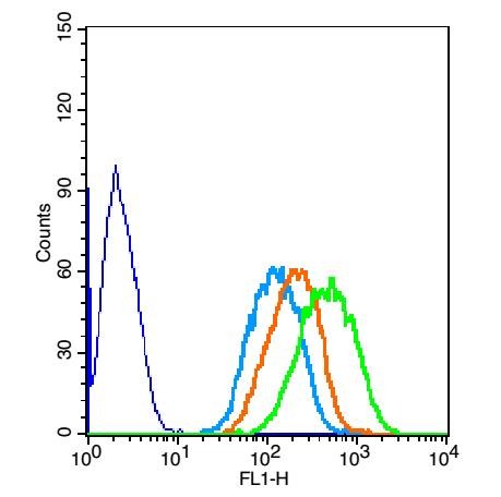 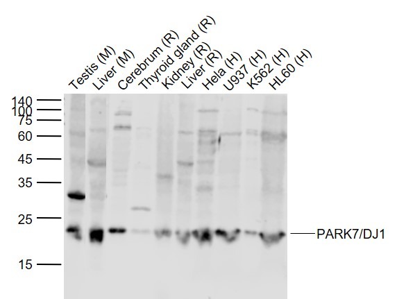 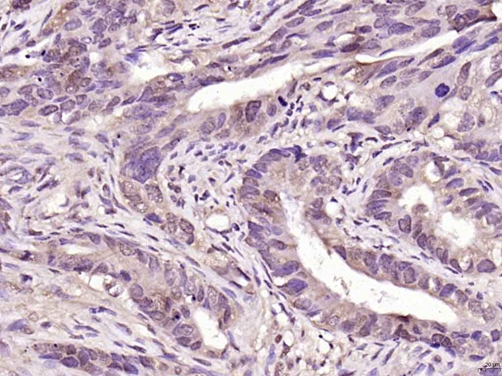 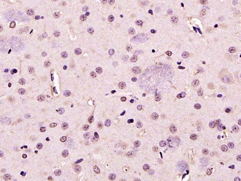 |
| 应用 | FCMIFIHC-FrIHC-PWB |
| 推荐剂量 | WB: 1:500-2000; IHC-P: 1:100-500; IHC-Fr: 1:100-500; IF: 1:100-500; FCM: 0.2μg/Test |
| 抗体种类 | Polyclonal |
| 宿主来源 | Rabbit |
| 亚细胞定位 | Cytoplasm. Nucleus. Mitochondrion. Under normal conditions, located predominantly in the cytoplasm and, to a lesser extent, in the nucleus and mitochondrion. Translocates to the mitochondrion and subsequently to the nucleus in response to oxidative stress and exerts an increased cytoprotective effect against oxidative damage. Detected in tau inclusions in brains from neurodegenerative disease patients. |
| 组织特异性 | Highly expressed in pancreas, kidney, skeletal muscle, liver, testis and heart. Detected at slightly lower levels in placenta and brain. Detected in astrocytes, Sertoli cells, spermatogonia, spermatids and spermatozoa. |
| 构建方式 | Polyclonal Antibody |
| 纯化方式 | Protein A purified |
| 性状 | Liquid |
| 缓冲液 | 0.01M TBS (pH7.4) with 1% BSA, 0.02% Proclin300 and 50% Glycerol. |
| 浓度 | 1 mg/mL |
| 研究背景 | PARK7/DJ1 is a ubiquitously expressed protein involved in various cellular processes including cell proliferation, RNA-binding, and oxidative stress. The protein has been found to colocalize within a subset of pathologic tau inclusions in a diverse group of neurodegenerative disorders known as tauopathies (Rizzu et al. 2004). Defects in PARK7/DJ1 are the cause of autosomal recessive early-onset Parkinson's disease 7 (PARK7). Parkinson's disease (PD) is a complex, multifactorial disorder that typically manifests after the age of 50 years. The disease is characterized by bradykinesia, resting tremor, muscular rigidity and postural instability. The pathology involves the loss of dopaminergic neurons in the substantia nigra and the presence of Lewy bodies (intraneuronal accumulations of aggregated proteins), in surviving neurons in various areas of the brain. PARK7 is characterized by onset before 40 years and slow progression. It has also been suggested that PARK7/DJ1 is a mitogen dependent oncogene product involved in Ras related signal transduction pathways. |
| 免疫原 | KLH conjugated synthetic peptide: human CAP1 |
| 抗原种属 | Human |
| 基因名称 | PARK7 |
| 基因ID | |
| 蛋白名称 | Parkinson disease protein 7 |
| Uniprot ID | |
| 研究领域 | Small G proteins,DNA / RNA binding,Oxidative stress,Parkin / PARK,Regulators |
| 功能 | Protects cells against oxidative stress and cell death. Plays a role in regulating expression or stability of the mitochondrial uncoupling proteins SLC25A14 and SLC25A27 in dopaminergic neurons of the substantia nigra pars compacta and attenuates the oxidative stress induced by calcium entry into the neurons via L-type channels during pacemaking. Eliminates hydrogen peroxide and protects cells against hydrogen peroxide-induced cell death. May act as an atypical peroxiredoxin-like peroxidase that scavenges hydrogen peroxide. Following removal of a C-terminal peptide, displays protease activity and enhanced cytoprotective action against oxidative stress-induced apoptosis. Stabilizes NFE2L2 by preventing its association with KEAP1 and its subsequent ubiquitination. Binds to OTUD7B and inhibits its deubiquitinating activity. Enhances RELA nuclear translocation. Binds to a number of mRNAs containing multiple copies of GG or CC motifs and partially inhibits their translation but dissociates following oxidative stress. Required for correct mitochondrial morphology and function and for autophagy of dysfunctional mitochondria. Regulates astrocyte inflammatory responses. Acts as a positive regulator of androgen receptor-dependent transcription. Prevents aggregation of SNCA. Plays a role in fertilization. Has no proteolytic activity. Has cell-growth promoting activity and transforming activity. May function as a redox-sensitive chaperone. |
| 分子量 | Theoretical: 20 kDa. Actual: 22 kDa. |
| 储存方式 | Store at -20°C or -80°C for 12 months. Avoid repeated freeze-thaw cycles. |
| 运输方式 | Shipping with blue ice. |
 嗨!有任何问题?点我咨询
版权所有©2015-2026 TargetMol Chemicals Inc.保留所有权利.
沪ICP备20019793号-4 | 沪公网安备 31010602006700号 | 沪(静)应急管危经许[2024]203441