

购物车
 您的购物车当前为空


 还可以
还可以Anti-TIE2 Polyclonal Antibody 是一种 Rabbit 抗体,靶向 TIE2。Anti-TIE2 Polyclonal Antibody 可用于 FCM, ICC/IF。
还可以别名 Tie-2, Tie2, TEK tyrosine kinase, endothelial, STK1, Hyk, Cd202b, AA517024
Anti-TIE2 Polyclonal Antibody 是一种 Rabbit 抗体,靶向 TIE2。Anti-TIE2 Polyclonal Antibody 可用于 FCM, ICC/IF。
| 规格 | 价格 | 库存 | 数量 |
|---|---|---|---|
| 50 μL | ¥ 1,165 | 5日内发货 | |
| 100 μL | ¥ 1,960 | 5日内发货 | |
| 200 μL | ¥ 2,785 | 5日内发货 |
TargetMol的所有产品仅用作科学研究或药证申报,不能被用于人体,我们不向个人提供产品和服务。请您遵守承诺用途,不得违反法律法规规定用于任何其他用途。
| 产品描述 | Anti-TIE2 Polyclonal Antibody is a Rabbit antibody targeting TIE2. Anti-TIE2 Polyclonal Antibody can be used in FCM, ICC/IF. |
| 别名 | Tie-2, Tie2, TEK tyrosine kinase, endothelial, STK1, Hyk, Cd202b, AA517024 |
| Ig Type | IgG |
| 反应种属 | Human (predicted:Mouse,Rat,Pig,Cow,Horse) |
| 验证活性 | 1. Tissue/cell: A549 cell; 4% Paraformaldehyde-fixed; Triton X-100 at room temperature for 20 min; Blocking buffer (normal goat serum) at 37°C for 20 min; Antibody incubation with (TIE2) polyclonal Antibody, Unconjugated (TMAB-01839) 1:100, 90 minutes at 37°C; followed by a FITC conjugated Goat Anti-Rabbit IgG antibody at 37°C for 90 minutes, DAPI (blue) was used to stain the cell nucleus. 2. Blank control (Black line): HUVEC (Black). Primary Antibody (green line): Rabbit Anti-TIE2 antibody (TMAB-01839) Dilution: 1 μg/10^6 cells; Isotype Control Antibody (orange line): Rabbit IgG. Secondary Antibody (white blue line): Goat anti-rabbit IgG-AF647 Dilution: 1 μg/test. Protocol The cells were fixed with 4% PFA (10 min at room temperature) and then permeabilized with 0.1% PBST for 20 min at room temperature. The cells were then incubated in 5% BSA to block non-specific protein-protein interactions for 30 min at room temperature. Cells stained with Primary Antibody for 30 min at room temperature. The secondary antibody used for 40 min at room temperature. 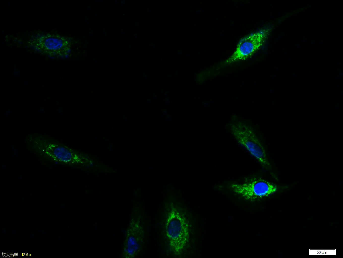 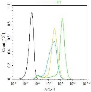 |
| 应用 | FCMICC/IF |
| 推荐剂量 | FCM=3 μg/Test; ICC/IF=1:100-500 |
| 抗体种类 | Polyclonal |
| 宿主来源 | Rabbit |
| 亚细胞定位 | Cell membrane; Single-pass type I membrane protein. Cell junction. Cell junction, focal adhesion. Cytoplasm, cytoskeleton. Secreted. Note=Recruited to cell-cell contacts in quiescent endothelial cells. Colocalizes with the actin cytoskeleton and at actin stress fibers during cell spreading. Recruited to the lower surface of migrating cells, especially the rear end of the cell. Proteolytic processing gives rise to a soluble extracellular domain that is secreted. |
| 组织特异性 | Predominantly expressed in endothelial cells and their progenitors, the angioblasts. Has been directly found in placenta and lung, with a lower level in umbilical vein endothelial cells, brain and kidney. |
| 构建方式 | Polyclonal Antibody |
| 纯化方式 | Protein A purified |
| 性状 | Liquid |
| 缓冲液 | 0.01M TBS (pH7.4) with 1% BSA, 0.02% Proclin300 and 50% Glycerol. |
| 浓度 | 1 mg/mL |
| 研究背景 | The TEK receptor tyrosine kinase is expressed almost exclusively in endothelial cells in mice, rats, and humans. This receptor possesses a unique extracellular domain containing 2 immunoglobulin-like loops separated by 3 epidermal growth factor-like repeats that are connected to 3 fibronectin type III-like repeats. The ligand for the receptor is angiopoietin-1. Defects in TEK are associated with inherited venous malformations; the TEK signaling pathway appears to be critical for endothelial cell-smooth muscle cell communication in venous morphogenesis.TEK is closely related to the TIE receptor tyrosine kinase. |
| 免疫原 | KLH conjugated synthetic peptide: human Tie2 |
| 抗原种属 | Human |
| 基因名称 | TEK |
| 基因ID | |
| 蛋白名称 | Angiopoietin-1 receptor |
| Uniprot ID | |
| 研究领域 | Endothelial Markers,Receptor Tyrosine Kinases,Angiogenesis and vasculogenesis,Endothelium,Endothelial Cells,Endothelial Cell Markers,Growth factor receptors |
| 功能 | This protein is a protein tyrosine-kinase transmembrane receptor for angiopoietin 1. It may constitute the earliest mammalian endothelial cell lineage marker. Probably regulates endothelial cell proliferation, differentiation and guides the proper patterning of endothelial cells during blood vessel formation. |
| 分子量 | Theoretical: 124 kDa. |
| 储存方式 | Store at -20°C or -80°C for 12 months. Avoid repeated freeze-thaw cycles. |
| 运输方式 | Shipping with blue ice. |
 嗨!有任何问题?点我咨询
版权所有©2015-2026 TargetMol Chemicals Inc.保留所有权利.
沪ICP备20019793号-4 | 沪公网安备 31010602006700号 | 沪(静)应急管危经许[2024]203441