

购物车
 您的购物车当前为空


 还可以
还可以Anti-FUBP1 Antibody (1H129) 是一种 Rabbit 抗体,靶向 FUBP1。Anti-FUBP1 Antibody (1H129) 可用于 FCM,ICC/IF,IHC,WB。
还可以别名 hDH V, FUSE-binding protein 1, FUBP 1, FBP, Far upstream element-binding protein 1, DNA helicase V
Anti-FUBP1 Antibody (1H129) 是一种 Rabbit 抗体,靶向 FUBP1。Anti-FUBP1 Antibody (1H129) 可用于 FCM,ICC/IF,IHC,WB。
| 规格 | 价格 | 库存 | 数量 |
|---|---|---|---|
| 50 μL | ¥ 1,480 | 5日内发货 | |
| 100 μL | ¥ 2,495 | 5日内发货 |
TargetMol的所有产品仅用作科学研究或药证申报,不能被用于人体,我们不向个人提供产品和服务。请您遵守承诺用途,不得违反法律法规规定用于任何其他用途。
| 产品描述 | Anti-FUBP1 Antibody (1H129) is a Rabbit antibody targeting FUBP1. Anti-FUBP1 Antibody (1H129) can be used in FCM,ICC/IF,IHC,WB. |
| 别名 | hDH V, FUSE-binding protein 1, FUBP 1, FBP, Far upstream element-binding protein 1, DNA helicase V |
| Ig Type | IgG |
| 克隆号 | 1H129 |
| 反应种属 | Human,Mouse,Rat |
| 验证活性 | 1. Western blot analysis of FUBP1 on different lysates using anti-FUBP1 antibody at 1/1,000 dilution. Positive control: Lane 1: Hela, Lane 2: Raji. 2. Immunohistochemical analysis of paraffin-embedded mouse pancreas tissue using anti-FUBP1 antibody. Counter stained with hematoxylin. 3. Immunohistochemical analysis of paraffin-embedded human breast carcinoma tissue using anti-FUBP1 antibody. Counter stained with hematoxylin. 4. Immunohistochemical analysis of paraffin-embedded human pancreas tissue using anti-FUBP1 antibody. Counter stained with hematoxylin. 5. Immunohistochemical analysis of paraffin-embedded mouse brain tissue using anti-FUBP1 antibody. Counter stained with hematoxylin. 6. Immunohistochemical analysis of paraffin-embedded human tonsil tissue using anti-FUBP1 antibody. Counter stained with hematoxylin. 7. Immunohistochemical analysis of paraffin-embedded mouse stomach tissue using anti-FUBP1 antibody. Counter stained with hematoxylin. 8. ICC staining FUBP1 in Hela cells (green). The nuclear counter stain is DAPI (blue). Cells were fixed in paraformaldehyde, permeabilised with 0.25% Triton X100/PBS. 9. ICC staining FUBP1 in MCF-7 cells (green). The nuclear counter stain is DAPI (blue). Cells were fixed in paraformaldehyde, permeabilised with 0.25% Triton X100/PBS. 10. ICC staining FUBP1 in HepG2 cells (green). The nuclear counter stain is DAPI (blue). Cells were fixed in paraformaldehyde, permeabilised with 0.25% Triton X100/PBS. 11. Flow cytometric analysis of Jurakt cells with FUBP1 antibody at 1/50 dilution (red) compared with an unlabelled control (cells without incubation with primary antibody; black). Alexa Fluor 488-conjugated goat anti rabbit IgG was used as the secondary antibody. 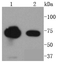 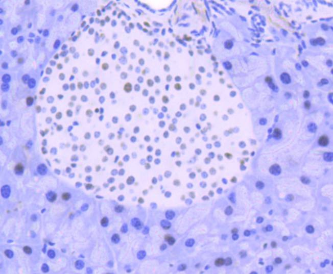 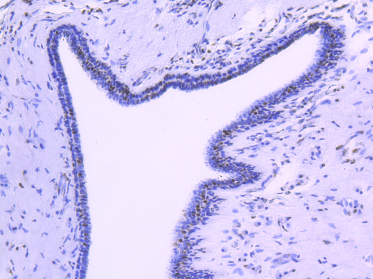  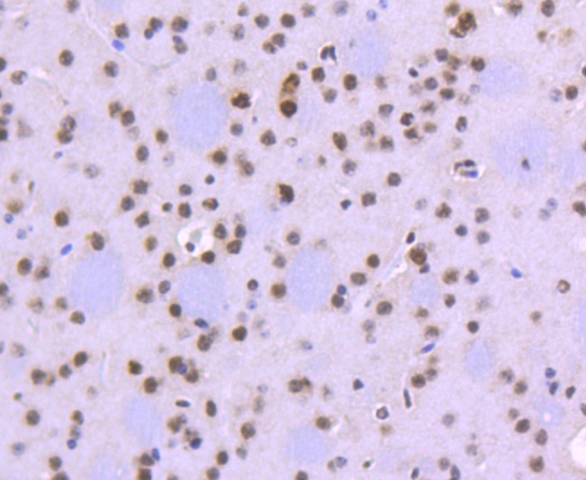 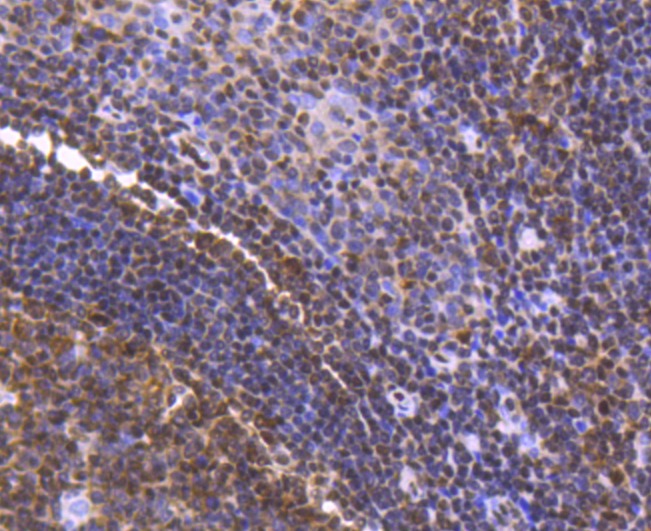 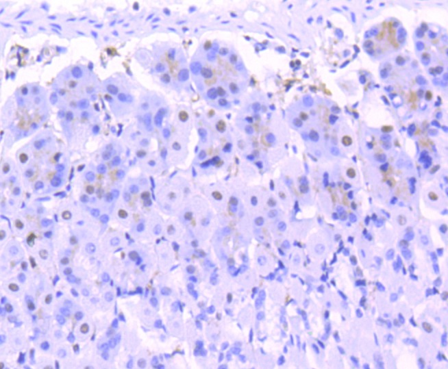 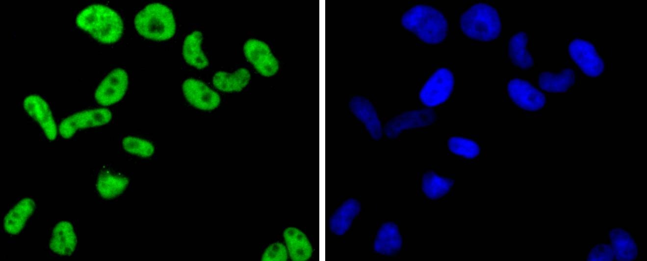 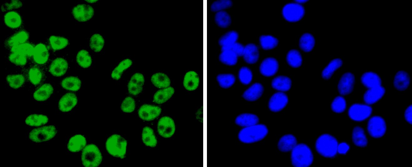 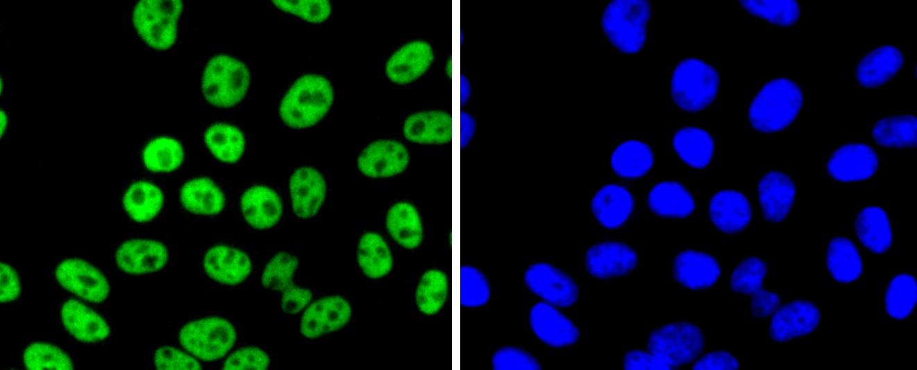 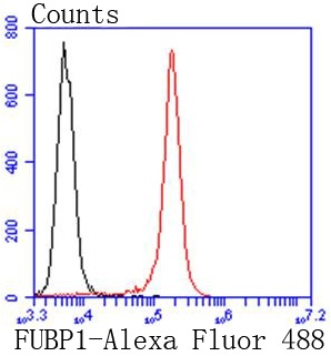 |
| 应用 | FCMICC/IFIHCWB |
| 推荐剂量 | WB: 1:1000-2000; IHC: 1:50-200; ICC/IF: 1:100-500; FCM: 1:50-100 |
| 抗体种类 | Monoclonal |
| 宿主来源 | Rabbit |
| 构建方式 | Recombinant Antibody |
| 纯化方式 | ProA affinity purified |
| 性状 | Liquid |
| 缓冲液 | 1*TBS (pH7.4), 1%BSA, 40%Glycerol. Preservative: 0.05% Sodium Azide. |
| 研究背景 | Activation of FUSE, the far upstream element, is required for the proper ex-pression of the mammalian gene c-Myc in undifferentiated cells. The binding of FBP1 (FUSE-binding protein or far upstream element-binding protein) to FUSE is necessary for c-Myc expression, indicating that FBP1 functions as a growth-dependent regulator of c-Myc expression. Isolated from proliferating HL-60 cells, FBP1 (FBP), FBP2 and FBP3 comprise a family of single-stranded DNA-binding proteins that specifically bind to FUSE elements. The FBP transcription factors share a conserved central DNA-binding domain and show significant homology in their carboxyl-terminal activation domains. Expression of FBP1 is detected in undifferentiated cells and is substantially decreased following cellular differentiation. |
| 偶联 | Unconjugated |
| 免疫原 | Recombinant Protein |
| Uniprot ID |
| 分子量 | Theoretical: 74 kDa. |
| 储存方式 | Store at -20°C or -80°C for 12 months. Avoid repeated freeze-thaw cycles. |
| 运输方式 | Shipping with blue ice. |
 嗨!有任何问题?点我咨询
版权所有©2015-2026 TargetMol Chemicals Inc.保留所有权利.
沪ICP备20019793号-4 | 沪公网安备 31010602006700号 | 沪(静)应急管危经许[2024]203441