

购物车
 您的购物车当前为空


 还可以
还可以Anti-ITGB1 Polyclonal Antibody 是一种 Rabbit 抗体,靶向 ITGB1。Anti-ITGB1 Polyclonal Antibody 可用于 FCM, IF, IHC-Fr, IHC-P, WB。
还可以别名 VLA-β, VLA-BETA, VLAB, MSK12, MDF2, integrin, β1 (fibronectin receptor, βpolypeptide, antigen CD29 includes MDF2, MSK12), integrin, beta 1 (fibronectin receptor, beta polypeptide, antigen CD29 includes MDF2, MSK12), Integrin β1, GPIIA, FNRB, CD29
Anti-ITGB1 Polyclonal Antibody 是一种 Rabbit 抗体,靶向 ITGB1。Anti-ITGB1 Polyclonal Antibody 可用于 FCM, IF, IHC-Fr, IHC-P, WB。
| 规格 | 价格 | 库存 | 数量 |
|---|---|---|---|
| 50 μL | ¥ 1,175 | 5日内发货 | |
| 100 μL | ¥ 1,975 | 5日内发货 | |
| 200 μL | ¥ 2,795 | 5日内发货 |
TargetMol的所有产品仅用作科学研究或药证申报,不能被用于人体,我们不向个人提供产品和服务。请您遵守承诺用途,不得违反法律法规规定用于任何其他用途。
| 产品描述 | Anti-ITGB1 Polyclonal Antibody is a Rabbit antibody targeting ITGB1. Anti-ITGB1 Polyclonal Antibody can be used in FCM, IF, IHC-Fr, IHC-P, WB. |
| 别名 | VLA-β, VLA-BETA, VLAB, MSK12, MDF2, integrin, β1 (fibronectin receptor, βpolypeptide, antigen CD29 includes MDF2, MSK12), integrin, beta 1 (fibronectin receptor, beta polypeptide, antigen CD29 includes MDF2, MSK12), Integrin β1, GPIIA, FNRB, CD29 |
| Ig Type | IgG |
| 反应种属 | Human,Mouse,Rat (predicted:Chicken,Dog,Pig,Cow,Horse,Rabbit,Sheep) |
| 验证活性 | 1. Blank control (Black line): HUVEc (Black). Primary Antibody (green line): Rabbit Anti-Integrin beta 1 antibody (TMAB-00996) Dilution: 3 μg/10^6 cells; Isotype Control Antibody (orange line): Rabbit IgG-PE. Protocol The cells were fixed with 4% PFA (10 min at room temperature) and then were incubated in 5% BSA to block non-specific protein-protein interactions for 30 min at room temperature. Cells stained with Primary Antibody for 30 min at room temperature. 2. Paraformaldehyde-fixed, paraffin embedded (Rat brain); Antigen retrieval by boiling in sodium citrate buffer (pH6.0) for 15 min; Block endogenous peroxidase by 3% hydrogen peroxide for 20 min; Blocking buffer (normal goat serum) at 37°C for 30 min; Antibody incubation with (Integrin beta 1) Polyclonal Antibody, Unconjugated (TMAB-00996) at 1:500 overnight at 4°C, followed by a conjugated secondary for 20 min and DAB staining. 3. Paraformaldehyde-fixed, paraffin embedded (Rat heart); Antigen retrieval by boiling in sodium citrate buffer (pH6.0) for 15 min; Block endogenous peroxidase by 3% hydrogen peroxide for 20 min; Blocking buffer (normal goat serum) at 37°C for 30 min; Antibody incubation with (Integrin beta 1) Polyclonal Antibody, Unconjugated (TMAB-00996) at 1:400 overnight at 4°C, followed by operating according to SP Kit (Rabbit) instructionsand DAB staining. 4. Blank control: HUVEC cells (blue). Primary Antibody: Rabbit Anti-CD29 antibody (TMAB-00996), Dilution: 1 μg in 100 μL 1X PBS containing 0.5% BSA; Isotype Control Antibody: Rabbit Igg (orange),used under the same conditions); Secondary Antibody: Goat anti-rabbit IgG-Pe (white blue), Dilution: 1:200 in 1 X PBS containing 0.5% BSA. Protocol The cells were fixed with 2% paraformaldehyde (10 min). Primary antibody (TMAB-00996, 1 μg/1x10^6 cells) were incubated for 30 min on the ice, followed by 1 X PBS containing 0.5% BSA + 10% goat serum (15 min) to block non-specific protein-protein interactions. Then the Goat Anti-rabbit IgG/PE antibody was added into the blocking buffer mentioned above to react with the primary antibody at 1/200 dilution for 30 min on ice. 5. Blank control (blue line): Hela (blue). Primary Antibody (green line): Rabbit Anti-Integrin beta 1 antibody (TMAB-00996) Dilution: 1 μg/10^6 cells; Isotype Control Antibody (orange line): Rabbit IgG. Secondary Antibody (white blue line): Goat anti-rabbit IgG-PE Dilution: 1 μg/test. Protocol The cells were fixed with 70% methanol (Overnight at 4°C) and then permeabilized with 90% ice-cold methanol for 20 min at-20°C. Cells stained with Primary Antibody for 30 min at room temperature. The cells were then incubated in 1 X PBS/2% BSA/10% goat serum to block non-specific protein-protein interactions followed by the antibody for 15 min at room temperature. The secondary antibody used for 40 min at room temperature. 6. Sample: Lane 1: A431 (Human) Cell Lysate at 30 μg Lane 2: U-87MG (Human) Cell Lysate at 30 μg Lane 3: Ear (Rat) Lysate at 40 μg Lane 4: Ear (Mouse) Lysate at 40 μg Lane 5: Bladder (Mouse) Lysate at 40 μg Primary: Anti-Integrin beta 1 (TMAB-00996) at 1/1000 dilution Secondary: IRDye800CW Goat Anti-Rabbit IgG at 1/20000 dilution Predicted band size: 130 kDa Observed band size: 130 kDa 7. Paraformaldehyde-fixed, paraffin embedded (Rat liver); Antigen retrieval by boiling in sodium citrate buffer (pH6.0) for 15 min; Block endogenous peroxidase by 3% hydrogen peroxide for 20 min; Blocking buffer (normal goat serum) at 37°C for 30 min; Antibody incubation with (Integrin beta 1) Polyclonal Antibody, Unconjugated (TMAB-00996) at 1:400 overnight at 4°C, followed by operating according to SP Kit (Rabbit) instructionsand DAB staining. 8. Paraformaldehyde-fixed, paraffin embedded (Mouse brain); Antigen retrieval by boiling in sodium citrate buffer (pH6.0) for 15 min; Block endogenous peroxidase by 3% hydrogen peroxide for 20 min; Blocking buffer (normal goat serum) at 37°C for 30 min; Antibody incubation with (Integrin beta 1) Polyclonal Antibody, Unconjugated (TMAB-00996) at 1:400 overnight at 4°C, followed by operating according to SP Kit (Rabbit) instructions and DAB staining. 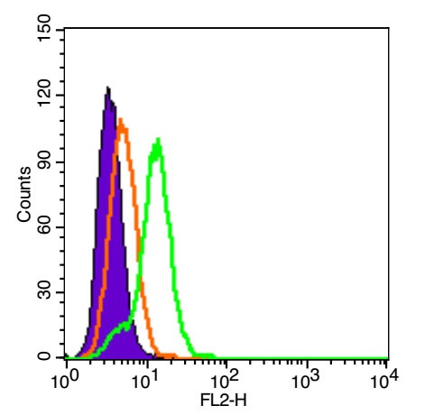 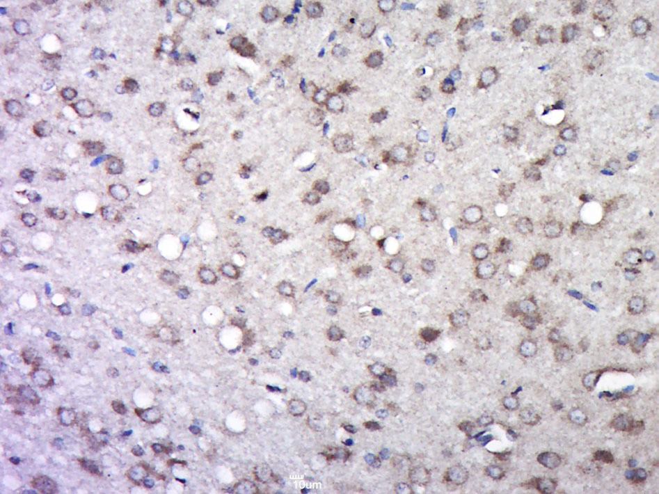 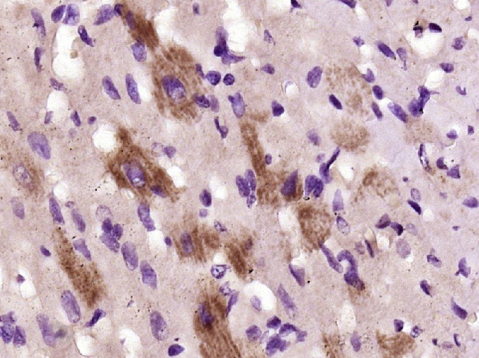 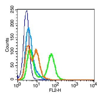 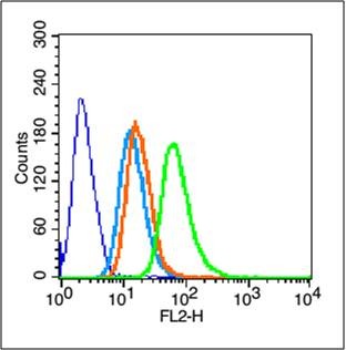 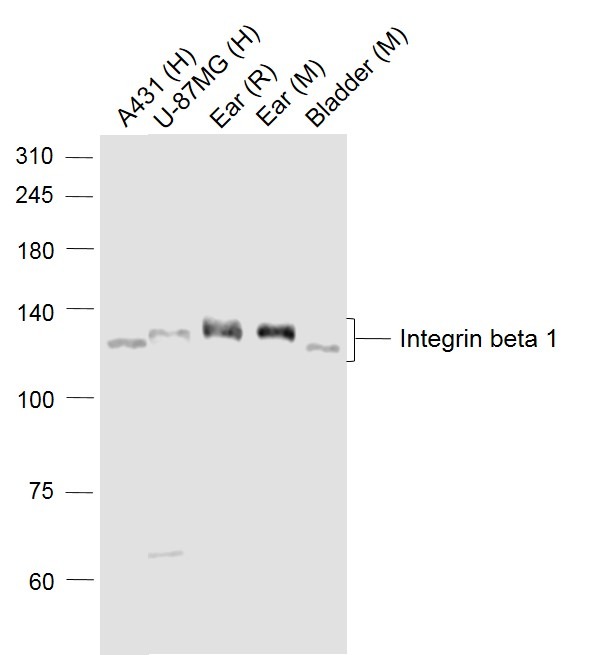 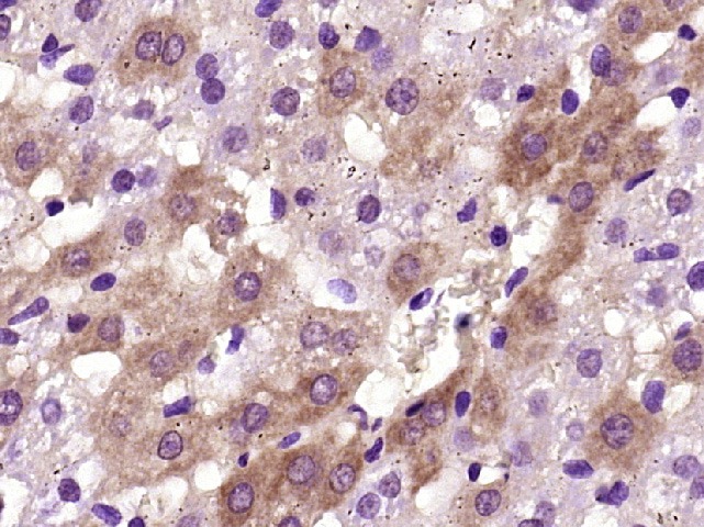 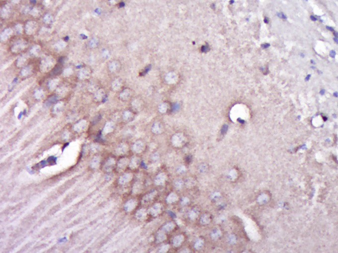 |
| 应用 | FCMIFIHC-FrIHC-PWB |
| 推荐剂量 | WB: 1:500-2000; IHC-P: 1:100-500; IHC-Fr: 1:100-500; IF: 1:100-500; FCM: 1μg/Test |
| 抗体种类 | Polyclonal |
| 宿主来源 | Rabbit |
| 亚细胞定位 | Cell membrane; Single-pass type I membrane protein. Melanosome. Cleavage furrow. Note=Isoform beta-1B does not localize to focal adhesions. Highly enriched in stage I melanosomes. Located on plasma membrane of neuroblastoma NMB7 cells. In a lung cancer cell line, in prometaphase and metaphase, localizes diffusely at the membrane and in afew intracellular vesicles. In early telophase, detected mainly on the matrix-facing side of the cells. By mid-telophase, concentrated to the ingressing cleavage furrow, mainly to the basal side of the furrow. In late telophase, concentrated to the extending protrusions formed at the opposite ends of the spreading daughter cells, in vesicles at the base of the lamellipodia formed by the separating daughter cells. |
| 组织特异性 | Isoform beta-1A is widely expressed, other isoforms are generally coexpressed with a more restricted distribution. Isoform beta-1B is expressed in skin, liver, skeletal muscle, cardiac muscle, placenta, umbilical vein endothelial cells, neuroblastoma cell |
| 构建方式 | Polyclonal Antibody |
| 纯化方式 | Protein A purified |
| 性状 | Liquid |
| 缓冲液 | 0.01M TBS (pH7.4) with 1% BSA, 0.02% Proclin300 and 50% Glycerol. |
| 浓度 | 1 mg/mL |
| 研究背景 | Integrins alpha-1/beta-1, alpha-2/beta-1, alpha-10/beta-1 and alpha-11/beta-1 are receptors for collagen. Integrins alpha-1/beta-1 and alpha-2/beta-2 recognize the proline-hydroxylated sequence G-F-P-G-E-R in collagen. Integrins alpha-2/beta-1, alpha-3/beta-1, alpha-4/beta-1, alpha-5/beta-1, alpha-8/beta-1, alpha-10/beta-1, alpha-11/beta-1 and alpha-V/beta-1 are receptors for fibronectin. Alpha-4/beta-1 recognizes one or more domains within the alternatively spliced CS-1 and CS-5 regions of fibronectin. Integrin alpha-5/beta-1 is a receptor for fibrinogen. Integrin alpha-1/beta-1, alpha-2/beta-1, alpha-6/beta-1 and alpha-7/beta-1 are receptors for lamimin. Integrin alpha-4/beta-1 is a receptor for VCAM1. It recognizes the sequence Q-I-D-S in VCAM1. Integrin alpha-9/beta-1 is a receptor for VCAM1, cytotactin and osteopontin. It recognizes the sequence A-E-I-D-G-I-E-L in cytotactin. Integrin alpha-3/beta-1 is a receptor for epiligrin, thrombospondin and CSPG4. Alpha-3/beta-1 may mediate with LGALS3 the stimulation by CSPG4 of endothelial cells migration. Integrin alpha-V/beta-1 is a receptor for vitronectin. Beta-1 integrins recognize the sequence R-G-D in a wide array of ligands. Isoform beta-1B interferes with isoform beta-1A resulting in a dominant negative effect on cell adhesion and migration (in vitro). In case of HIV-1 infection, the interaction with extracellular viral Tat protein seems to enhance angiogenesis in Kaposi's sarcoma lesions. |
| 免疫原 | KLH conjugated synthetic peptide: human Integrin beta 1 |
| 抗原种属 | Human |
| 基因名称 | ITGB1 |
| 基因ID | |
| 蛋白名称 | Integrin beta-1 |
| Uniprot ID | |
| 研究领域 | Beta |
| 功能 | Integrins alpha-1/beta-1, alpha-2/beta-1, alpha-10/beta-1 and alpha-11/beta-1 are receptors for collagen. Integrins alpha-1/beta-1 and alpha-2/beta-2 recognize the proline-hydroxylated sequence G-F-P-G-E-R in collagen. Integrins alpha-2/beta-1, alpha-3/beta-1, alpha-4/beta-1, alpha-5/beta-1, alpha-8/beta-1, alpha-10/beta-1, alpha-11/beta-1 and alpha-V/beta-1 are receptors for fibronectin. Alpha-4/beta-1 recognizes one or more domains within the alternatively spliced CS-1 and CS-5 regions of fibronectin. Integrin alpha-5/beta-1 is a receptor for fibrinogen. Integrin alpha-1/beta-1, alpha-2/beta-1, alpha-6/beta-1 and alpha-7/beta-1 are receptors for lamimin. Integrin alpha-4/beta-1 is a receptor for VCAM1. It recognizes the sequence Q-I-D-S in VCAM1. Integrin alpha-9/beta-1 is a receptor for VCAM1, cytotactin and osteopontin. It recognizes the sequence A-E-I-D-G-I-E-L in cytotactin. Integrin alpha-3/beta-1 is a receptor for epiligrin, thrombospondin and CSPG4. Alpha-3/beta-1 may mediate with LGALS3 the stimulation by CSPG4 of endothelial cells migration. Integrin alpha-V/beta-1 is a receptor for vitronectin. Beta-1 integrins recognize the sequence R-G-D in a wide array of ligands. Isoform beta-1B interferes with isoform beta-1A resulting in a dominant negative effect on cell adhesion and migration (in vitro). In case of HIV-1 infection, the interaction with extracellular viral Tat protein seems to enhance angiogenesis in Kaposi's sarcoma lesions. |
| 分子量 | Theoretical: 88 kDa. Actual: 130 kDa. |
| 储存方式 | Store at -20°C or -80°C for 12 months. Avoid repeated freeze-thaw cycles. |
| 运输方式 | Shipping with blue ice. |
 嗨!有任何问题?点我咨询
版权所有©2015-2026 TargetMol Chemicals Inc.保留所有权利.
沪ICP备20019793号-4 | 沪公网安备 31010602006700号 | 沪(静)应急管危经许[2024]203441