

购物车
 您的购物车当前为空


 还可以
还可以Anti-CIDEC Polyclonal Antibody 是一种 Rabbit 抗体,靶向 CIDEC。Anti-CIDEC Polyclonal Antibody 可用于 FCM, ICC/IF, IF, IHC-Fr, IHC-P, WB。
还可以别名 FSP27, FLJ20871, Fat-specific protein FSP27 homolog, Fat specific protein 27, CIDEC, CIDE3, CIDE C, CIDE 3, Cell death-inducing DFFA-like effector protein C, Cell Death Inducing DFFA Like Effector C, Cell death activator CIDE-3, Cell Death Activator
Anti-CIDEC Polyclonal Antibody 是一种 Rabbit 抗体,靶向 CIDEC。Anti-CIDEC Polyclonal Antibody 可用于 FCM, ICC/IF, IF, IHC-Fr, IHC-P, WB。
| 规格 | 价格 | 库存 | 数量 |
|---|---|---|---|
| 50 μL | ¥ 1,170 | 5日内发货 | |
| 100 μL | ¥ 1,970 | 5日内发货 | |
| 200 μL | ¥ 2,785 | 5日内发货 |
TargetMol的所有产品仅用作科学研究或药证申报,不能被用于人体,我们不向个人提供产品和服务。请您遵守承诺用途,不得违反法律法规规定用于任何其他用途。
| 产品描述 | Anti-CIDEC Polyclonal Antibody is a Rabbit antibody targeting CIDEC. Anti-CIDEC Polyclonal Antibody can be used in FCM, ICC/IF, IF, IHC-Fr, IHC-P, WB. |
| 别名 | FSP27, FLJ20871, Fat-specific protein FSP27 homolog, Fat specific protein 27, CIDEC, CIDE3, CIDE C, CIDE 3, Cell death-inducing DFFA-like effector protein C, Cell Death Inducing DFFA Like Effector C, Cell death activator CIDE-3, Cell Death Activator |
| Ig Type | IgG |
| 反应种属 | Human,Mouse,Rat (predicted:Pig) |
| 验证活性 | 1. Paraformaldehyde-fixed, paraffin embedded (Rat brain); Antigen retrieval by boiling in sodium citrate buffer (pH6.0) for 15 min; Block endogenous peroxidase by 3% hydrogen peroxide for 20 min; Blocking buffer (normal goat serum) at 37°C for 30 min; Antibody incubation with (CIDEC) Polyclonal Antibody, Unconjugated (TMAB-00432) at 1:400 overnight at 4°C, followed by operating according to SP Kit (Rabbit) instructionsand DAB staining. 2. Tissue/cell: rat heart tissue; 4% Paraformaldehyde-fixed and paraffin-embedded; Antigen retrieval: citrate buffer (0.01M, pH6.0), Boiling bathing for 15 min; Block endogenous peroxidase by 3% Hydrogen peroxide for 30 min; Blocking buffer (normal goat serum) at 37°C for 20 min; Incubation: Anti-CIDEC Polyclonal Antibody, Unconjugated (TMAB-00432) 1:200, overnight at 4°C, followed by conjugation to the secondary antibody and DAb staining. 3. Tissue/cell: mouse stomach wall; 4% Paraformaldehyde-fixed and paraffin-embedded; Antigen retrieval: citrate buffer (0.01M, pH6.0), Boiling bathing for 15 min; Block endogenous peroxidase by 3% Hydrogen peroxide for 30 min; Blocking buffer (normal goat serum) at 37°C for 20 min; Incubation: Anti-CIDEC Polyclonal Antibody, Unconjugated (TMAB-00432) 1:200, overnight at 4°C, followed by conjugation to the secondary antibody and DAb staining. 4. Blank control: Mouse spleen. Primary Antibody (green line): Rabbit Anti-CIDEC antibody (TMAB-00432) Dilution: 2 μg/10^6 cells; Isotype Control Antibody (orange line): Rabbit IgG. Secondary Antibody: Goat anti-rabbit IgG-AF647 Dilution: 1 μg/test. Protocol The cells were fixed with 4% PFA (10 min at room temperature) and then permeabilized with 90% ice-cold methanol for 20 min at-20°C. The cells were then incubated in 5% BSA to block non-specific protein-protein interactions for 30 min at room temperature. Cells stained with Primary Antibody for 30 min at room temperature. The secondary antibody used for 40 min at room temperature. 5. Paraformaldehyde-fixed, paraffin embedded (rat breast); Antigen retrieval by boiling in sodium citrate buffer (pH6.0) for 15 min; Block endogenous peroxidase by 3% hydrogen peroxide for 20 min; Blocking buffer (normal goat serum) at 37°C for 30 min; Antibody incubation with (CIDEC) Polyclonal Antibody, Unconjugated (TMAB-00432) at 1:200 overnight at 4°C, followed by operating according to SP Kit (Rabbit) instructionsand DAB staining. 6. Paraformaldehyde-fixed, paraffin embedded (Mouse colon); Antigen retrieval by boiling in sodium citrate buffer (pH6.0) for 15 min; Block endogenous peroxidase by 3% hydrogen peroxide for 20 min; Blocking buffer (normal goat serum) at 37°C for 30 min; Antibody incubation with (CIDEC) Polyclonal Antibody, Unconjugated (TMAB-00432) at 1:200 overnight at 4°C, followed by operating according to SP Kit (Rabbit) instructionsand DAB staining. 7. Paraformaldehyde-fixed, paraffin embedded (rat colon); Antigen retrieval by boiling in sodium citrate buffer (pH6.0) for 15 min; Block endogenous peroxidase by 3% hydrogen peroxide for 20 min; Blocking buffer (normal goat serum) at 37°C for 30 min; Antibody incubation with (CIDEC) Polyclonal Antibody, Unconjugated (TMAB-00432) at 1:200 overnight at 4°C, followed by operating according to SP Kit (Rabbit) instructionsand DAB staining. 8. Paraformaldehyde-fixed, paraffin embedded (human gastric); Antigen retrieval by boiling in sodium citrate buffer (pH6.0) for 15 min; Block endogenous peroxidase by 3% hydrogen peroxide for 20 min; Blocking buffer (normal goat serum) at 37°C for 30 min; Antibody incubation with (CIDEC) Polyclonal Antibody, Unconjugated (TMAB-00432) at 1:200 overnight at 4°C, followed by operating according to SP Kit (Rabbit) instructionsand DAB staining. 9. Paraformaldehyde-fixed, paraffin embedded (rat brain); Antigen retrieval by boiling in sodium citrate buffer (pH6.0) for 15 min; Block endogenous peroxidase by 3% hydrogen peroxide for 20 min; Blocking buffer (normal goat serum) at 37°C for 30 min; Antibody incubation with (CIDEC) Polyclonal Antibody, Unconjugated (TMAB-00432) at 1:200 overnight at 4°C, followed by operating according to SP Kit (Rabbit) instructionsand DAB staining. 10. Paraformaldehyde-fixed, paraffin embedded (mouse brain); Antigen retrieval by boiling in sodium citrate buffer (pH6.0) for 15 min; Block endogenous peroxidase by 3% hydrogen peroxide for 20 min; Blocking buffer (normal goat serum) at 37°C for 30 min; Antibody incubation with (CIDEC) Polyclonal Antibody, Unconjugated (TMAB-00432) at 1:200 overnight at 4°C, followed by operating according to SP Kit (Rabbit) instructionsand DAB staining. 11. Paraformaldehyde-fixed, paraffin embedded (Human colon carcinoma); Antigen retrieval by boiling in sodium citrate buffer (pH6.0) for 15 min; Block endogenous peroxidase by 3% hydrogen peroxide for 20 min; Blocking buffer (normal goat serum) at 37°C for 30 min; Antibody incubation with (CIDEC) Polyclonal Antibody, Unconjugated (TMAB-00432) at 1:400 overnight at 4°C, followed by operating according to SP Kit (Rabbit) instructionsand DAB staining. 12. Blank control: A431. Primary Antibody (green line): Rabbit Anti-CIDEC antibody (TMAB-00432) Dilution: 1 μg/Test; Secondary Antibody: Goat anti-rabbit IgG-FITC Dilution: 0.5 μg/Test. Protocol The cells were fixed with 4% PFA (10 min at room temperature) and then permeabilized with 0.1% PBST for 20 min at room temperature. The cells were then incubated in 5% BSA to block non-specific protein-protein interactions for 30 min at room temperature. Cells stained with Primary Antibody for 30 min at room temperature. The secondary antibody used for 40 min at room temperature. 13. Sample: Lane 1: Adipose (Mouse) Lysate at 40 μg Lane 2: Breast (Mouse) Lysate at 40 μg Lane 3: Adipose (Rat) Lysate at 40 μg Primary: Anti-CIDEC (TMAB-00432) at 1/1000 dilution Secondary: IRDye800CW Goat Anti-Rabbit IgG at 1/20000 dilution Predicted band size: 27-30 kDa Observed band size: 27 kDa 14. Sample: Epididymis (Mouse) Lysate at 40 μg Primary: Anti-CIDEC (TMAB-00432) at 1/500 dilution Secondary: IRDye800CW Goat Anti-Rabbit IgG at 1/20000 dilution Predicted band size: 27 kDa Observed band size: 27 kDa 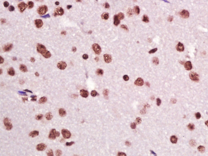 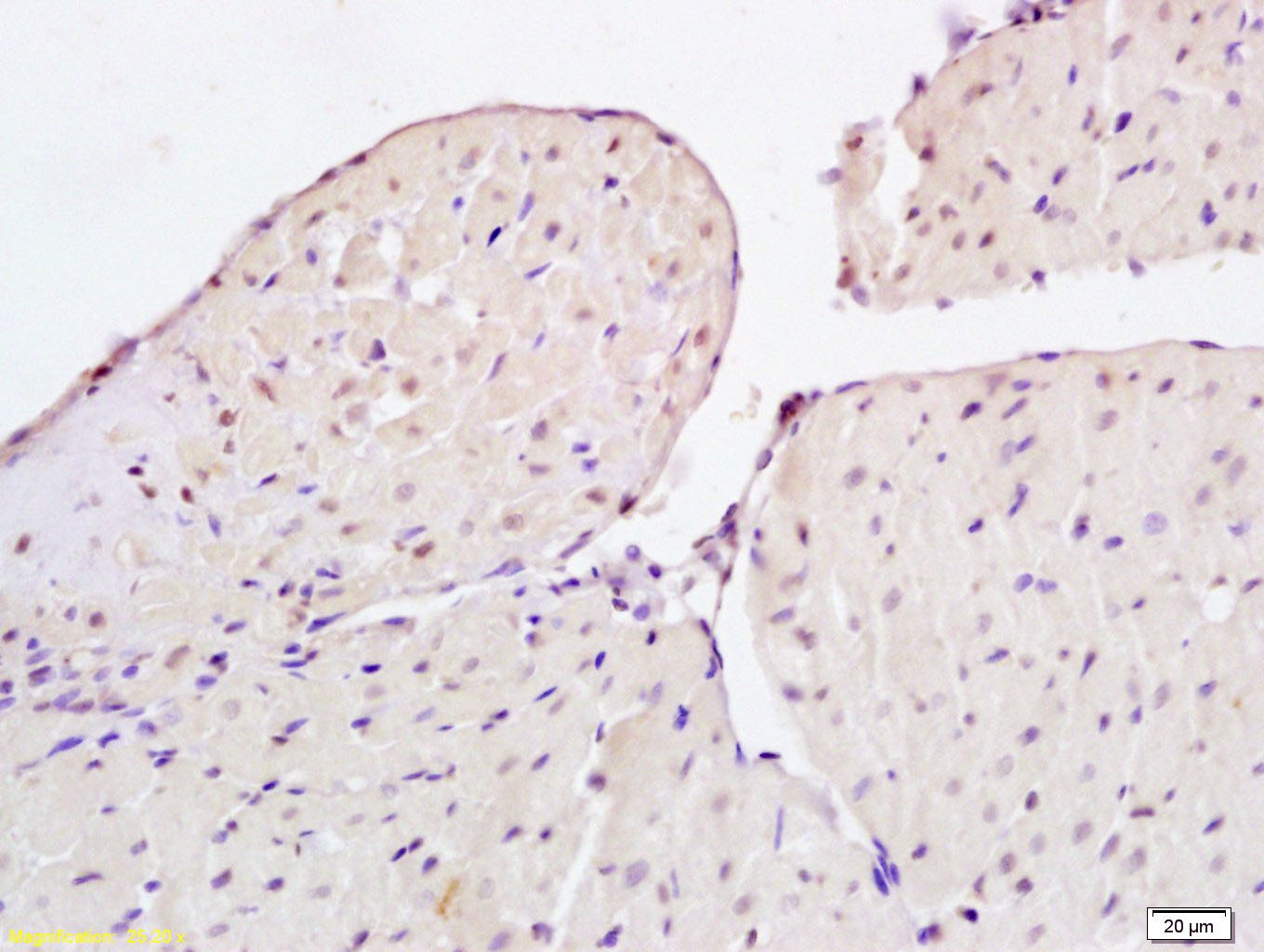 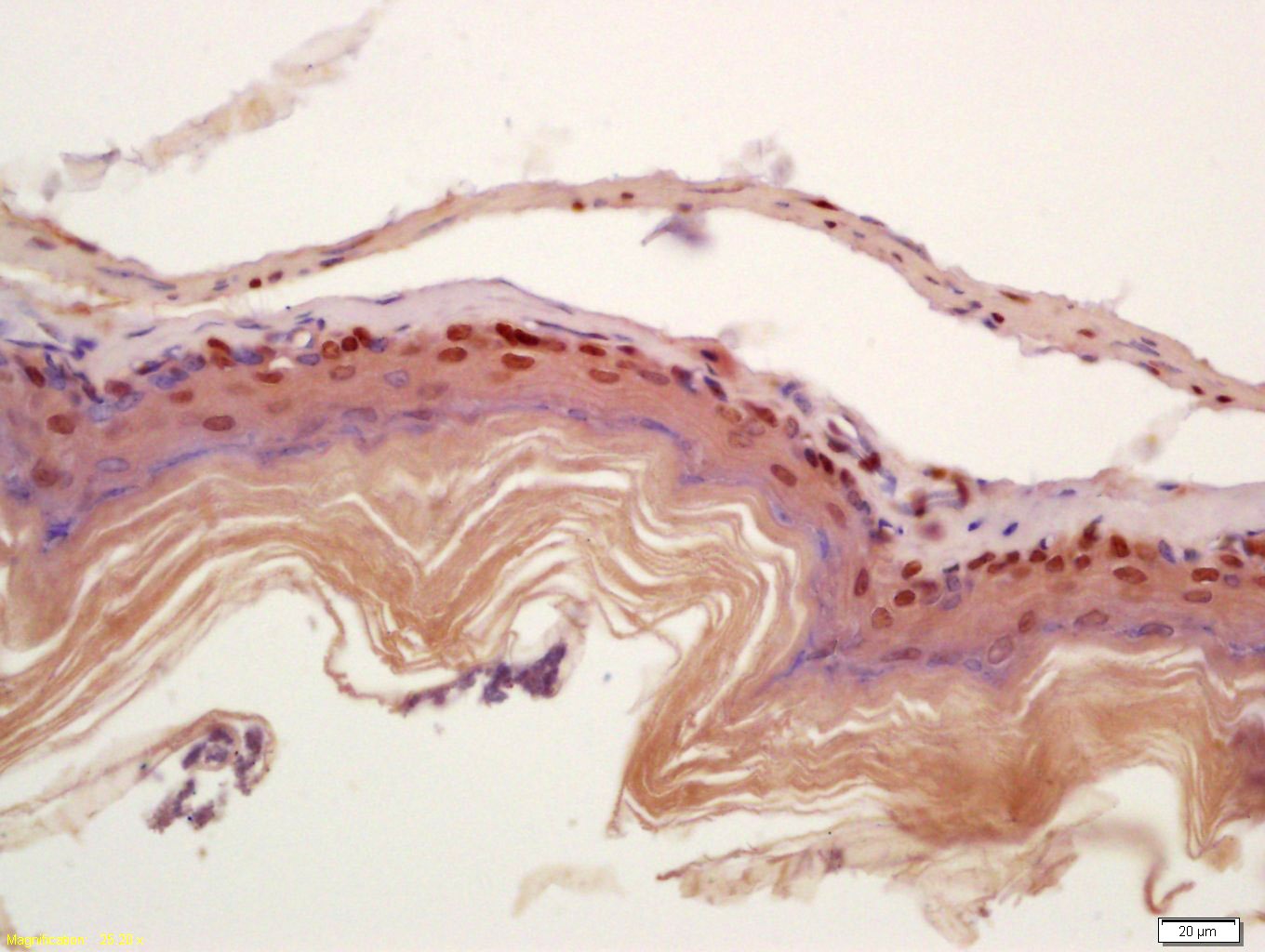 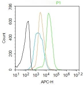 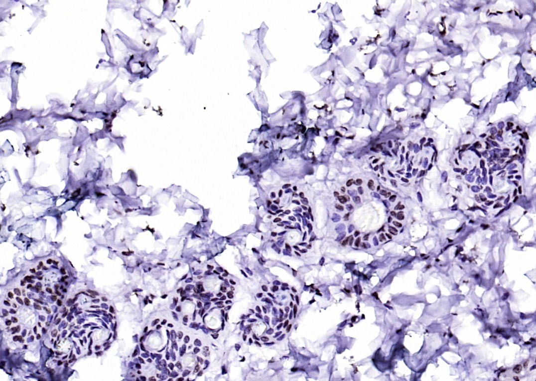 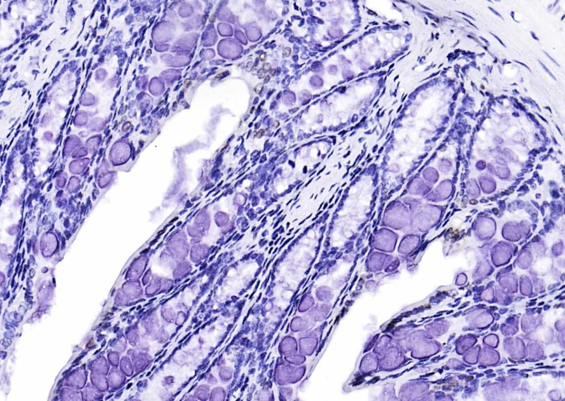 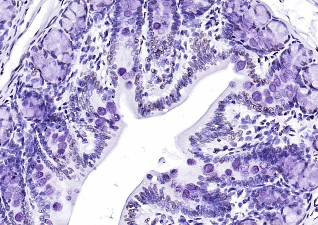 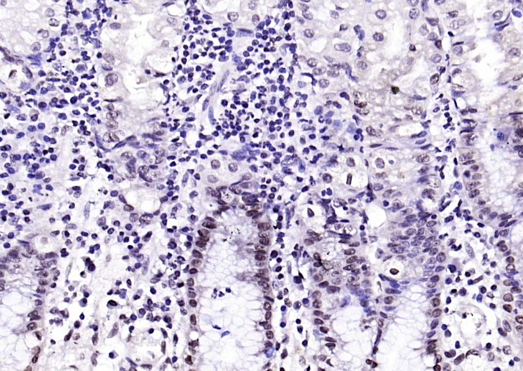 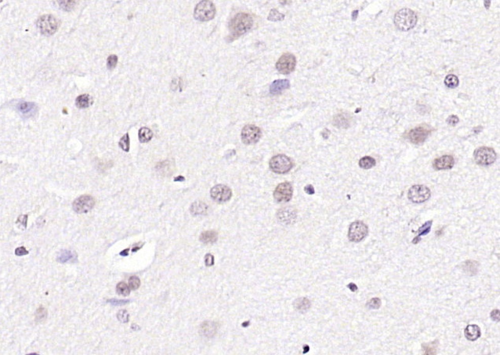 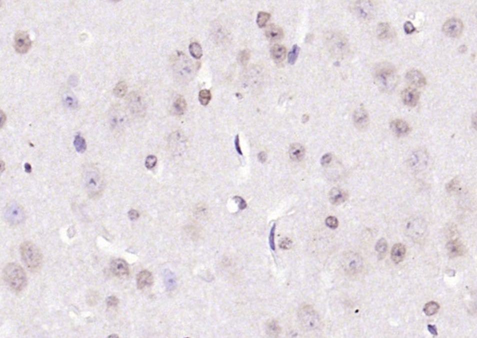 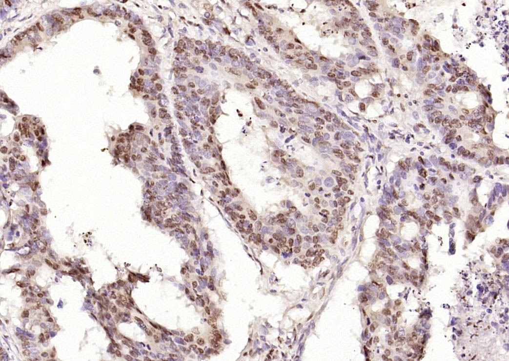 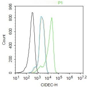 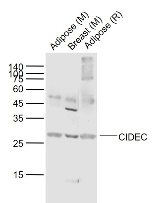 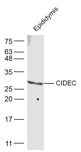 |
| 应用 | FCMICC/IFIFIHC-FrIHC-PWB |
| 推荐剂量 | FCM=1 μg/Test; ICC/IF=1:50-200; IF=1:100-500; IHC-Fr=1:100-500; IHC-P=1:100-500; WB=1:500-2000 |
| 抗体种类 | Polyclonal |
| 宿主来源 | Rabbit |
| 亚细胞定位 | Nucleus (By similarity). Endoplasmicreticulum (By similarity). Lipid droplet. Note=Diffuses quickly onlipid droplet surface, but becomes trapped and clustered at lipiddroplet contact sites, thereby enabling its rapid enrichment atlipid droplet contact sites. |
| 组织特异性 | Expressed mainly in adipose tissue, smallintestine, heart, colon and stomach and, at lower levels, in brain,kidney and liver. |
| 构建方式 | Polyclonal Antibody |
| 纯化方式 | Protein A purified |
| 性状 | Liquid |
| 缓冲液 | 0.01M TBS (pH7.4) with 1% BSA, 0.02% Proclin300 and 50% Glycerol. |
| 浓度 | 1 mg/mL |
| 研究背景 | This gene encodes a member of the cell death-inducing DNA fragmentation factor-like effector family. Members of this family play important roles in apoptosis. The encoded protein promotes lipid droplet formation in adipocytes and may mediate adipocyte apoptosis. This gene is regulated by insulin and its expression is positively correlated with insulin sensitivity. Mutations in this gene may contribute to insulin resistant diabetes. A pseudogene of this gene is located on the short arm of chromosome 3. Alternatively spliced transcript variants that encode different isoforms have been observed for this gene. [provided by RefSeq, Dec 2010]. Tissue specificity: Expressed mainly in small intestine, heart, colon and stomach and, at lower levels, in brain, kidney and liver. |
| 免疫原 | KLH conjugated synthetic peptide: human CIDEC |
| 抗原种属 | Human |
| 基因名称 | CIDEC |
| 基因ID | |
| 蛋白名称 | Cell death activator CIDE-3 |
| Uniprot ID | |
| 功能 | May act as a CEBPB coactivator in white adipose tissueto control the expression of a subset of CEBPB downstream targetgenes, including SOCS1, SOCS3, TGFB1, TGFBR1, ID2 and XDH (Bysimilarity). Binds to lipid droplets and regulates theirenlargement, thereby restricting lipolysis and favoring storage. Atfocal contact sites between lipid droplets, promotes directionalnet neutral lipid transfer from the smaller to larger lipiddroplets. The transfer direction may be driven by the internalpressure difference between the contacting lipid droplet pair. Whenoverexpressed in preadipocytes, induces apoptosis or increases cellsusceptibility to apoptosis induced by serum deprivation or TGFBtreatment. As mature adipocytes, that express high CIDEC levels,are quite resistant to apoptotic stimuli, the physiologicalsignificance of its role in apoptosis is unclear. |
| 分子量 | Theoretical: 27 kDa. Actual: 27 kDa. |
| 储存方式 | Store at -20°C or -80°C for 12 months. Avoid repeated freeze-thaw cycles. |
| 运输方式 | Shipping with blue ice. |
 嗨!有任何问题?点我咨询
版权所有©2015-2026 TargetMol Chemicals Inc.保留所有权利.
沪ICP备20019793号-4 | 沪公网安备 31010602006700号 | 沪(静)应急管危经许[2024]203441