

购物车
 您的购物车当前为空


 还可以
还可以Anti-BAX Polyclonal Antibody 2 是一种 Rabbit 抗体,靶向 BAX。Anti-BAX Polyclonal Antibody 2 可用于 FCM, ICC/IF, IF, IHC-Fr, IHC-P, WB。
还可以别名 Bcl-2-like protein 4, Bcl2-L-4, BCL2L4, Apoptosis regulator BAX
Anti-BAX Polyclonal Antibody 2 是一种 Rabbit 抗体,靶向 BAX。Anti-BAX Polyclonal Antibody 2 可用于 FCM, ICC/IF, IF, IHC-Fr, IHC-P, WB。
| 规格 | 价格 | 库存 | 数量 |
|---|---|---|---|
| 50 μL | ¥ 1,175 | 5日内发货 | |
| 100 μL | ¥ 1,960 | 5日内发货 | |
| 200 μL | ¥ 2,790 | 5日内发货 |
TargetMol的所有产品仅用作科学研究或药证申报,不能被用于人体,我们不向个人提供产品和服务。请您遵守承诺用途,不得违反法律法规规定用于任何其他用途。
| 产品描述 | Anti-BAX Polyclonal Antibody 2 is a Rabbit antibody targeting BAX. Anti-BAX Polyclonal Antibody 2 can be used in FCM, ICC/IF, IF, IHC-Fr, IHC-P, WB. |
| 别名 | Bcl-2-like protein 4, Bcl2-L-4, BCL2L4, Apoptosis regulator BAX |
| Ig Type | IgG |
| 反应种属 | Human,Mouse,Rat (predicted:Dog,Pig,Cow,Rabbit,Sheep) |
| 验证活性 | 1. Sample: Hela (Human) Cell Lysate at 30 μg Primary: Anti-Bax (TMAB-00188) at 1/1000 dilution Secondary: IRDye800CW Goat Anti-Rabbit IgG at 1/20000 dilution Predicted band size: 21 kDa Observed band size: 23 kDa 2. Paraformaldehyde-fixed, paraffin embedded (Human liver carcinoma); Antigen retrieval by boiling in sodium citrate buffer (pH6.0) for 15 min; Block endogenous peroxidase by 3% hydrogen peroxide for 20 min; Blocking buffer (normal goat serum) at 37°C for 30 min; Antibody incubation with (Bax) Polyclonal Antibody, Unconjugated (TMAB-00188) at 1:400 overnight at 4°C, followed by operating according to SP Kit (Rabbit) instructionsand DAB staining. 3. Paraformaldehyde-fixed, paraffin embedded (Rat brain); Antigen retrieval by boiling in sodium citrate buffer (pH6.0) for 15 min; Block endogenous peroxidase by 3% hydrogen peroxide for 20 min; Blocking buffer (normal goat serum) at 37°C for 30 min; Antibody incubation with (Bax) Polyclonal Antibody, Unconjugated (TMAB-00188) at 1:400 overnight at 4°C, followed by operating according to SP Kit (Rabbit) instructionsand DAB staining. 4. Overlay histogram showing HL 60 cells stained with TMAB-00188 (Green line). The cells were fixed with 90% methanol (5 min) and then permeabilized with 0.01M PBS-Tween for 20 min. The cells were then incubated in 1x PBS / 10% normal goat serum to block non-specific protein-protein interactions followed by the antibody (TMAB-00188,1 μg/1x10^6 cells) for 30 min at 22°C. The secondary antibody used was fluorescein isothiocyanate goat anti-rabbit IgG (H+L) (Brillant blue line) at 1/200 dilution for 30 min at 22°C. Isotype control antibody was rabbit IgG (polyclonal, Orange line) (1 μg/1x10^6 cells) used under the same conditions. Unlabelled sample (blue line) was also used as a control. Acquisition of 20,000 events were collected using a 20mW Argon ion laser (488nm) and 525/30 bandpass filter. 5. Sample: Lane 1: Testis (Mouse) Lysate at 40 μg Lane 2: Kidney (Rat) Lysate at 40 μg Lane 3: Testis (Rat) Lysate at 40 μg Primary: Anti-Bax (TMAB-00188) at 1/1000 dilution Secondary: IRDye800CW Goat Anti-Rabbit IgG at 1/20000 dilution Predicted band size: 21 kDa Observed band size: 21 kDa 6. Tissue/cell: SH-SY5Y cell; 4% Paraformaldehyde-fixed; Triton X-100 at room temperature for 20 min; Blocking buffer (normal goat serum) at 37°C for 20 min; Antibody incubation with (Bax) polyclonal Antibody, Unconjugated (TMAB-00188) 1:100, 90 minutes at 37°C; followed by a FITC conjugated Goat Anti-Rabbit IgG antibody at 37°C for 90 minutes, DAPI (blue) was used to stain the cell nucleus. 7. Tissue/cell: SH-SY5Y cell; 4% Paraformaldehyde-fixed; Triton X-100 at room temperature for 20 min; Blocking buffer (normal goat serum) at 37°C for 20 min; Antibody incubation with (Bax) polyclonal Antibody, Unconjugated (TMAB-00188) 1:100, 90 minutes at 37°C; followed by a FITC conjugated Goat Anti-Rabbit IgG antibody at 37°C for 90 minutes, DAPI (blue) was used to stain the cell nucleus. 8. Paraformaldehyde-fixed, paraffin embedded (Rat spinal cord); Antigen retrieval by boiling in sodium citrate buffer (pH6.0) for 15 min; Block endogenous peroxidase by 3% hydrogen peroxide for 20 min; Blocking buffer (normal goat serum) at 37°C for 30 min; Antibody incubation with (Bax) Polyclonal Antibody, Unconjugated (TMAB-00188) at 1:400 overnight at 4°C, followed by operating according to SP Kit (Rabbit) instructionsand DAB staining. 9. Sample: Cerebral cortex (Rat) Lysate at 40 μg Primary: Anti-Bax (TMAB-00188) at 1/1000 dilution Secondary: IRDye800CW Goat Anti-Rabbit IgG at 1/20000 dilution Predicted band size: 21 kDa Observed band size: 21 kDa 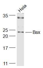 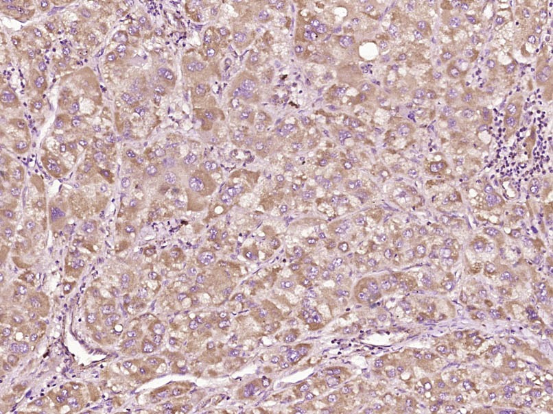 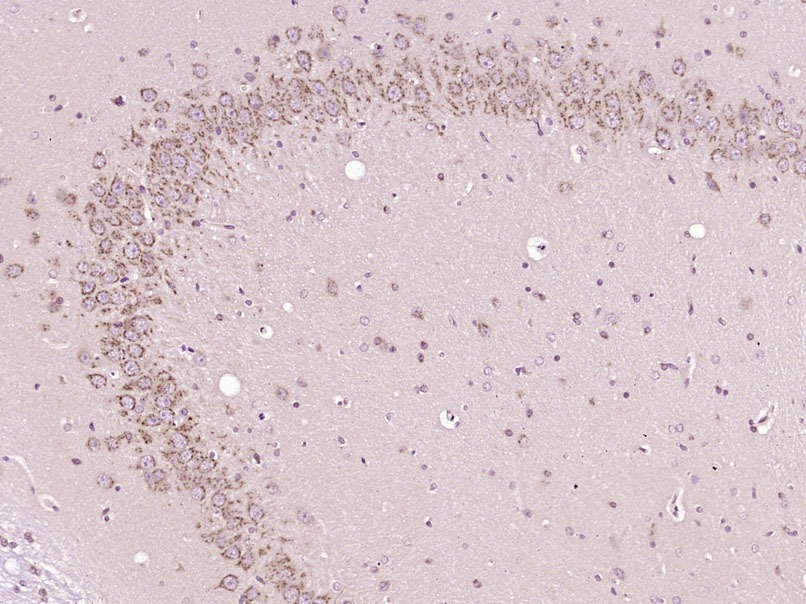 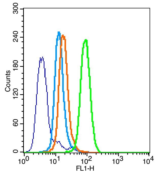 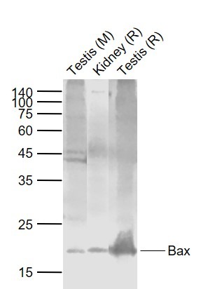 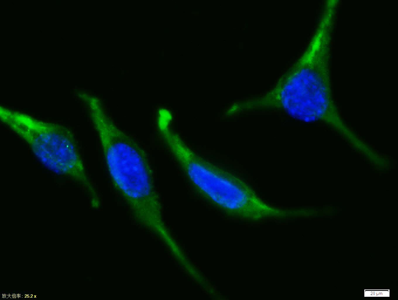 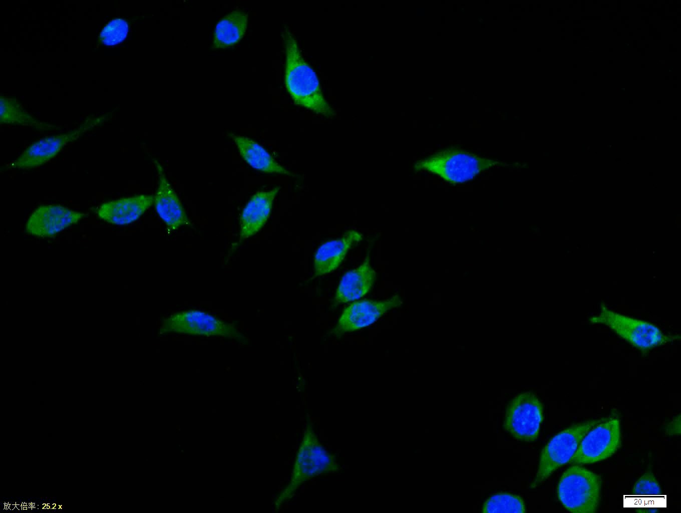  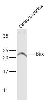 |
| 应用 | FCMICC/IFIFIHC-FrIHC-PWB |
| 推荐剂量 | FCM=1 μg/Test; ICC/IF=1:50-200; IF=1:100-500; IHC-Fr=1:100-500; IHC-P=1:100-500; WB=1:500-2000 |
| 抗体种类 | Polyclonal |
| 宿主来源 | Rabbit |
| 亚细胞定位 | Isoform Alpha: Mitochondrion membrane; Single-pass membrane protein. Cytoplasm. Note=Colocalizes with 14-3-3 proteins in the cytoplasm. Under stress conditions, undergoes a conformation change that causes release from JNK-phosphorylated 14-3-3 proteins and translocation to the mitochondrion membrane. Isoform Beta: Cytoplasm. Isoform Gamma: Cytoplasm. Isoform Delta: Cytoplasm (Potential). |
| 组织特异性 | Expressed in a wide variety of tissues. Isoform Psi is found in glial tumors. Isoform Alpha is expressed in spleen, breast, ovary, testis, colon and brain, and at low levels in skin and lung. Isoform Sigma is expressed in spleen, breast, ovary, testis, lu |
| 构建方式 | Polyclonal Antibody |
| 纯化方式 | Protein A purified |
| 性状 | Liquid |
| 缓冲液 | 0.01M TBS (pH7.4) with 1% BSA, 0.02% Proclin300 and 50% Glycerol. |
| 浓度 | 1 mg/mL |
| 研究背景 | The protein encoded by this gene belongs to the BCL2 protein family. BCL2 family members form hetero- or homodimers and act as anti- or pro-apoptotic regulators that are involved in a wide variety of cellular activities. This protein forms a heterodimer with BCL2, and functions as an apoptotic activator. This protein is reported to interact with, and increase the opening of, the mitochondrial voltage-dependent anion channel (VDAC), which leads to the loss in membrane potential and the release of cytochrome c. The expression of this gene is regulated by the tumor suppressor P53 and has been shown to be involved in P53-mediated apoptosis. Multiple alternatively spliced transcript variants, which encode different isoforms, have been reported for this gene. [provided by RefSeq, Jul 2008]. |
| 免疫原 | KLH conjugated synthetic peptide: human Bax |
| 抗原种属 | Human |
| 基因名称 | BAX |
| 基因ID | |
| 蛋白名称 | Apoptosis regulator BAX |
| Uniprot ID | |
| 研究领域 | Bcl 2 family,Metabolism,Bcl 2 family,Associated Proteins,Bcl2 Family,Apoptosis marker and proteins ELISA kits,Apoptosis |
| 功能 | Accelerates programmed cell death by binding to, and antagonizing the apoptosis repressor BCL2 or its adenovirus homolog E1B 19k protein. Under stress conditions, undergoes a conformation change that causes translocation to the mitochondrion membrane, leading to the release of cytochrome c that then triggers apoptosis. Promotes activation of CASP3, and thereby apoptosis. |
| 分子量 | Theoretical: 21 kDa. Actual: 20-23 kDa. |
| 储存方式 | Store at -20°C or -80°C for 12 months. Avoid repeated freeze-thaw cycles. |
| 运输方式 | Shipping with blue ice. |
 嗨!有任何问题?点我咨询
版权所有©2015-2026 TargetMol Chemicals Inc.保留所有权利.
沪ICP备20019793号-4 | 沪公网安备 31010602006700号 | 沪(静)应急管危经许[2024]203441