

购物车
 您的购物车当前为空


 还可以
还可以Anti-NR4A1 Antibody (7O127) 是一种 Rabbit 抗体,靶向 NR4A1。Anti-NR4A1 Antibody (7O127) 可用于 ICC/IF,IHC,WB。
还可以别名 Testicular receptor 3, ST-59, Orphan nuclear receptor TR3, Orphan nuclear receptor HMR, Nuclear receptor subfamily 4immunitygroup A member 1, Nuclear hormone receptor NUR/77 (Nur77), NR4A1, NAK1, HMR, GFRP1, Early response protein NAK1
Anti-NR4A1 Antibody (7O127) 是一种 Rabbit 抗体,靶向 NR4A1。Anti-NR4A1 Antibody (7O127) 可用于 ICC/IF,IHC,WB。
| 规格 | 价格 | 库存 | 数量 |
|---|---|---|---|
| 50 μL | ¥ 1,490 | 5日内发货 | |
| 100 μL | ¥ 2,490 | 5日内发货 |
TargetMol的所有产品仅用作科学研究或药证申报,不能被用于人体,我们不向个人提供产品和服务。请您遵守承诺用途,不得违反法律法规规定用于任何其他用途。
| 产品描述 | Anti-NR4A1 Antibody (7O127) is a Rabbit antibody targeting NR4A1. Anti-NR4A1 Antibody (7O127) can be used in ICC/IF,IHC,WB. |
| 别名 | Testicular receptor 3, ST-59, Orphan nuclear receptor TR3, Orphan nuclear receptor HMR, Nuclear receptor subfamily 4immunitygroup A member 1, Nuclear hormone receptor NUR/77 (Nur77), NR4A1, NAK1, HMR, GFRP1, Early response protein NAK1 |
| Ig Type | IgG |
| 克隆号 | 7O127 |
| 反应种属 | Human,Mouse,Rat |
| 验证活性 | 1. Western blot analysis of NUR77 on rat brain cells lysates using anti-NUR77 antibody at 1/500 dilution. 2. ICC staining of NUR77 in Hela cells (green). 4% PFA fixed cells 20 minutes, washed with PBS. Cells were probed with the primary antibody (49510, 1/50) overnight at 4℃ washed with PBS. CoraLite488 Goat anti-Rabbit lgG was used as the secondary antibody at 1/100 dilution. The nuclear counter stain is DAPI (blue). 3. ICC staining of NUR77 in 3T3-L1 cells (green). 4% PFA fixed cells 20 minutes, washed with PBS. Cells were probed with the primary antibody (49510, 1/50) overnight at 4℃ washed with PBS. CoraLite488 Goat anti-Rabbit lgG was used as the secondary antibody at 1/100 dilution. The nuclear counter stain is DAPI (blue). 4. Immunohistochemical analysis of paraffin-embedded human liver tissue with Rabbit anti-NUR77 antibody (ET1703-97) at 1/1,000 dilution. The section was pre-treated using heat mediated antigen retrieval with sodium citrate buffer (pH 6.0) for 2 minutes. The tissues were blocked in 1% BSA for 20 minutes at room temperature, washed with ddH2O and PBS, and then probed with the primary antibody at 1/1,000 dilution for 1 hour at room temperature. The detection was performed using an HRP conjugated compact polymer system. DAB was used as the chromogen. Tissues were counterstained with hematoxylin and mounted with DPX. 5. Immunohistochemical analysis of paraffin-embedded human colon carcinoma tissue with Rabbit anti-NUR77 antibody at 1/1,000 dilution. The section was pre-treated using heat mediated antigen retrieval with sodium citrate buffer (pH 6.0) for 2 minutes. The tissues were blocked in 1% BSA for 20 minutes at room temperature, washed with ddH2O and PBS, and then probed with the primary antibody at 1/1,000 dilution for 1 hour at room temperature. The detection was performed using an HRP conjugated compact polymer system. DAB was used as the chromogen. Tissues were counterstained with hematoxylin and mounted with DPX. 6. Immunohistochemical analysis of paraffin-embedded human breast tissue using anti-NUR77 antibody. The section was pre-treated using heat mediated antigen retrieval with Tris-EDTA buffer (pH 8.0-8.4) for 20 minutes.The tissues were blocked in 5% BSA for 30 minutes at room temperature, washed with ddH2O and PBS, and then probed with the primary antibody (1/50) for 30 minutes at room temperature. The detection was performed using an HRP conjugated compact polymer system. DAB was used as the chromogen. Tissues were counterstained with hematoxylin and mounted with DPX. 7. Immunohistochemical analysis of paraffin-embedded human breast tissue using anti-NUR77 antibody. The section was pre-treated using heat mediated antigen retrieval with Tris-EDTA buffer (pH 8.0-8.4) for 20 minutes.The tissues were blocked in 5% BSA for 30 minutes at room temperature, washed with ddH2O and PBS, and then probed with the primary antibody (1/50) for 30 minutes at room temperature. The detection was performed using an HRP conjugated compact polymer system. DAB was used as the chromogen. Tissues were counterstained with hematoxylin and mounted with DPX. 8. Immunohistochemical analysis of paraffin-embedded mouse ovarian tissue using anti-NUR77 antibody. The section was pre-treated using heat mediated antigen retrieval with Tris-EDTA buffer (pH 8.0-8.4) for 20 minutes.The tissues were blocked in 5% BSA for 30 minutes at room temperature, washed with ddH2O and PBS, and then probed with the primary antibody (1/50) for 30 minutes at room temperature. The detection was performed using an HRP conjugated compact polymer system. DAB was used as the chromogen. Tissues were counterstained with hematoxylin and mounted with DPX. 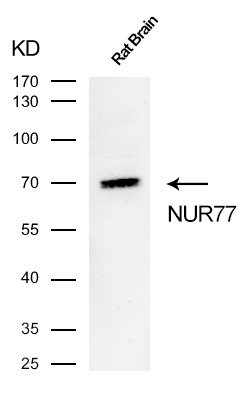 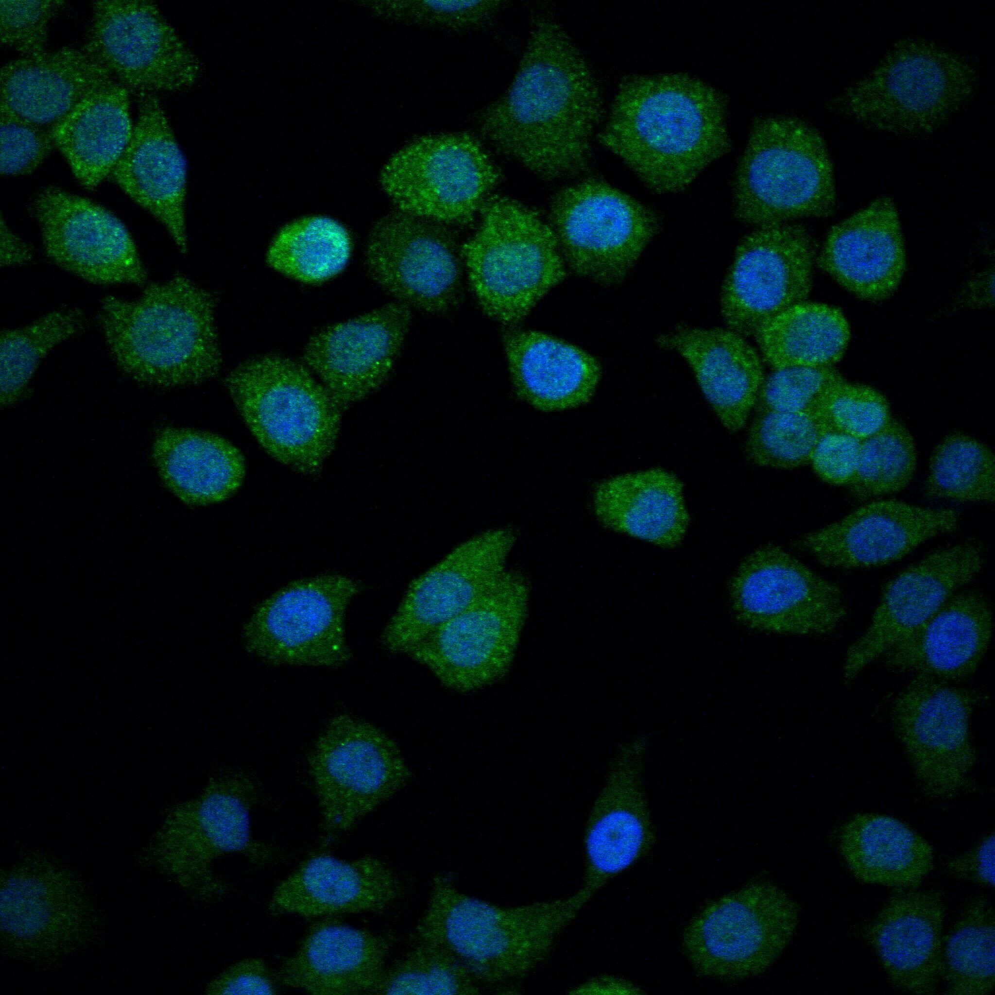 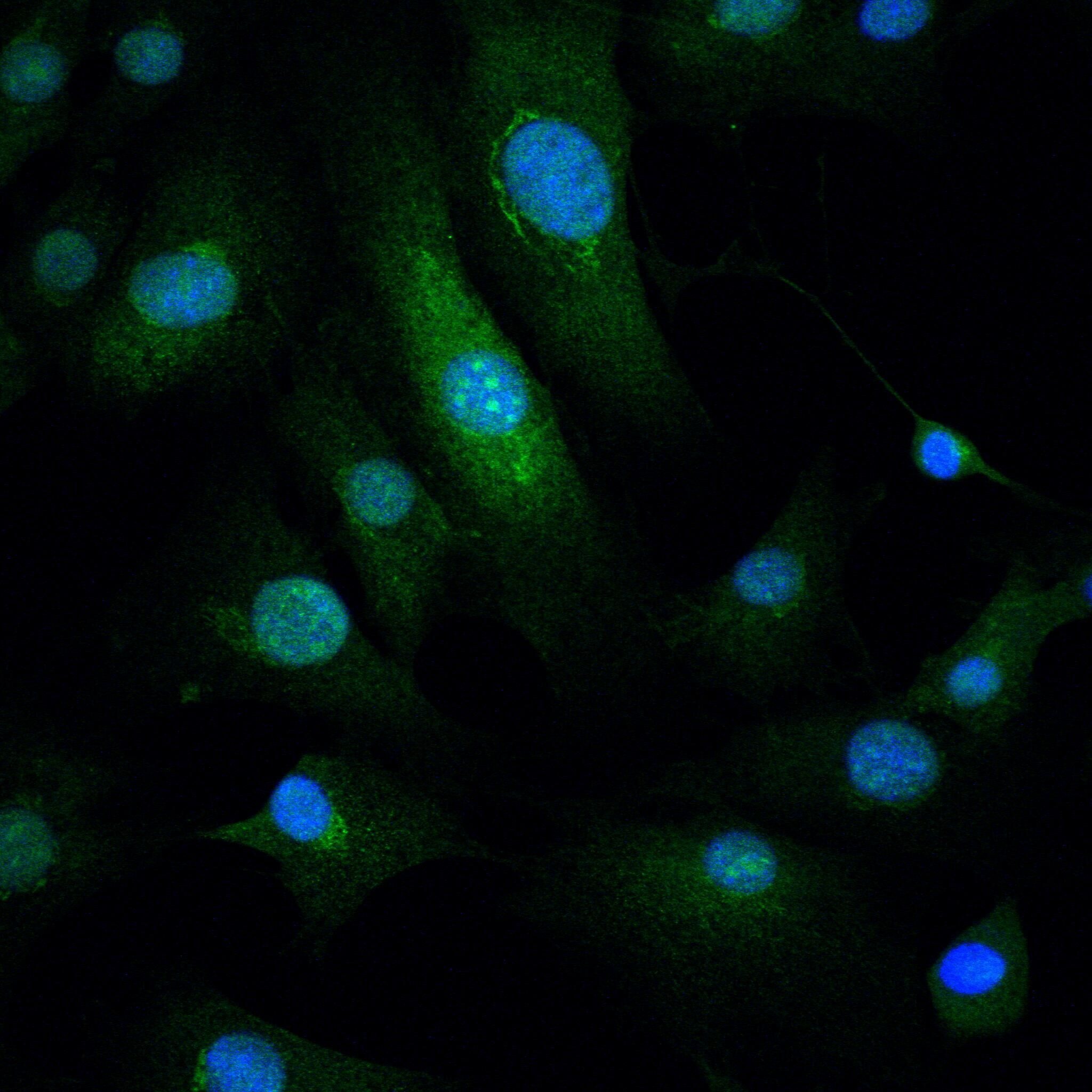  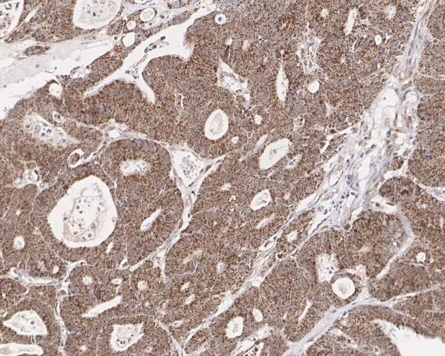 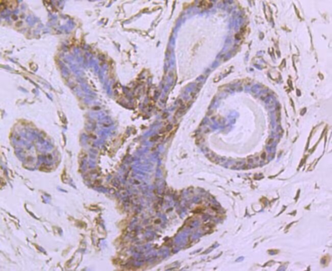 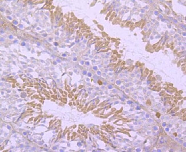 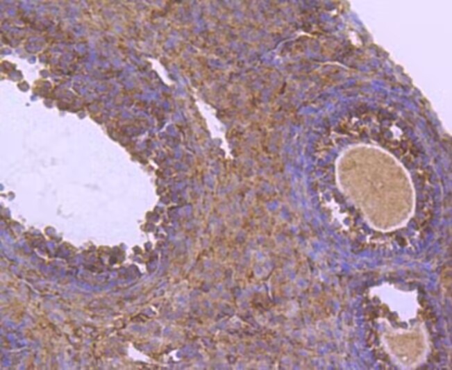 |
| 应用 | ICC/IFIHCWB |
| 推荐剂量 | WB: 1:500-1000; IHC: 1:50-200; ICC/IF: 1:50-200 |
| 抗体种类 | Monoclonal |
| 宿主来源 | Rabbit |
| 构建方式 | Recombinant Antibody |
| 纯化方式 | ProA affinity purified |
| 性状 | Liquid |
| 缓冲液 | 1*TBS (pH7.4), 0.05% BSA, 40% Glycerol. Preservative: 0.05% Sodium Azide. |
| 研究背景 | Nurr1 (Nur-related factor 1) and Nur77 (also designated NGFI-B) encode orphan nuclear receptors which may comprise an additional subfamily within the nuclear receptor superfamily. The rat and human homologs of mouse Nurr1 are designated RNR1 and NOT, respectively. Both Nurr1 and Nur77 are growth factor inducible immediate early response genes. Induction of both Nurr1 and Nur77 is seen after membrane depolarization while only Nur77 induction is seen with NGF stimulation. JunD acts as a mediator for Nur77. An increase in Nur77 expression is seen in activated T cells during G0 to G1 transition and throughout the G1 phase. In addition to its function as an immediate early gene, Nur77 may play a role in TCR-mediated apoptosis. Cyclosporin A, a potent immunosuppressant, has been shown to inhibit the ability of Nur77 to bind DNA. A dominant negative form of Nur77 can protect T cell hybridomas from activation-induced apoptosis. However, the absolute requirement of Nur77 for TCR-mediated apoptosis is still under debate. |
| 偶联 | Unconjugated |
| 免疫原 | Synthetic peptide within Human NUR77 aa 10-49 / 598 |
| 抗原种属 | Human |
| Uniprot ID |
| 分子量 | Theoretical: 64 kDa. |
| 储存方式 | Store at -20°C or -80°C for 12 months. Avoid repeated freeze-thaw cycles. |
| 运输方式 | Shipping with blue ice. |
 嗨!有任何问题?点我咨询
版权所有©2015-2026 TargetMol Chemicals Inc.保留所有权利.
沪ICP备20019793号-4 | 沪公网安备 31010602006700号 | 沪(静)应急管危经许[2024]203441