

购物车
 您的购物车当前为空


 还可以
还可以Anti-FUT4 Polyclonal Antibody 是一种 Rabbit 抗体,靶向 FUT4。Anti-FUT4 Polyclonal Antibody 可用于 FCM,IF,IHC-Fr,IHC-P,WB。
还可以别名 SSEA-1, SSEA1, LeX, Galactoside 3-L-fucosyltransferase, FUTIV, FucT-IV), FUC-TIV, Fucosyltransferase IV(Fuc-TIV, Fucosyltransferase 4, FCT3A, ELFT, ELAM-1 ligand fucosyltransferase, EC:2.4.1.152, CD15, alpha-(1, 3)-fucosyltransferase 4, 4-galactosyl-N-acetylglucosaminide 3-alpha-L-fucosyltransferase
Anti-FUT4 Polyclonal Antibody 是一种 Rabbit 抗体,靶向 FUT4。Anti-FUT4 Polyclonal Antibody 可用于 FCM,IF,IHC-Fr,IHC-P,WB。
| 规格 | 价格 | 库存 | 数量 |
|---|---|---|---|
| 50 μL | ¥ 1,165 | 5日内发货 | |
| 100 μL | ¥ 1,965 | 5日内发货 | |
| 200 μL | ¥ 2,780 | 5日内发货 |
TargetMol的所有产品仅用作科学研究或药证申报,不能被用于人体,我们不向个人提供产品和服务。请您遵守承诺用途,不得违反法律法规规定用于任何其他用途。
| 产品描述 | Anti-FUT4 Polyclonal Antibody is a Rabbit antibody targeting FUT4. Anti-FUT4 Polyclonal Antibody can be used in FCM,IF,IHC-Fr,IHC-P,WB. |
| 别名 | SSEA-1, SSEA1, LeX, Galactoside 3-L-fucosyltransferase, FUTIV, FucT-IV), FUC-TIV, Fucosyltransferase IV(Fuc-TIV, Fucosyltransferase 4, FCT3A, ELFT, ELAM-1 ligand fucosyltransferase, EC:2.4.1.152, CD15, alpha-(1, 3)-fucosyltransferase 4, 4-galactosyl-N-acetylglucosaminide 3-alpha-L-fucosyltransferase |
| Ig Type | IgG |
| 反应种属 | Human,Mouse (predicted:Rat) |
| 验证活性 | 1. Paraformaldehyde-fixed, paraffin embedded (Human lung cancer); Antigen retrieval by boiling in sodium citrate buffer (pH6.0) for 15 min; Block endogenous peroxidase by 3% hydrogen peroxide for 20 min; Blocking buffer (normal goat serum) at 37°C for 30 min; Antibody incubation with (FUT4) Polyclonal Antibody, Unconjugated (TMAB-00722) at 1:400 overnight at 4°C, followed by operating according to SP Kit (Rabbit) instructionsand DAB staining. 2. Overlay histogram showing HL 60 cells stained with TMAB-00722 (Green line). The cells were fixed with 90% methanol (5 min) and then permeabilized with 0.01M PBS-Tween for 20 min. The cells were then incubated in 1x PBS / 10% normal goat serum to block non-specific protein-protein interactions followed by the antibody (TMAB-00722,1 μg/1x10^6 cells) for 30 min at 22°C. The secondary antibody used was fluorescein isothiocyanate goat anti-rabbit IgG (H+L) (Brillant blue line) at 1/200 dilution for 30 min at 22°C. Isotype control antibody was rabbit IgG (polyclonal, Orange line) (1 μg/1x10^6 cells) used under the same conditions. Unlabelled sample (blue line) was also used as a control. Acquisition of 20,000 events were collected using a 20mW Argon ion laser (488nm) and 525/30 bandpass filter. 3. Blank control: Mouse spleen. Primary Antibody (green line): Rabbit Anti-FUT4/FITC Conjugated antibody (TMAB-00722-FITC) Dilution: 1 μg/10^6 cells; Isotype Control Antibody (orange line): Rabbit IgG-FITC. Protocol The cells were fixed with 4% PFA (10 min at room temperature) and then permeabilized with 0.1% PBST for 20 min at-20°C. The cells were then incubated in 5% BSA to block non-specific protein-protein interactions for 30 min at room temperature. The cells were stained with Primary Antibody for 30 min at room temperature. 4. Blank control: HL-60. Primary Antibody (green line): Rabbit Anti-FUT4 (TMAB-00722) Dilution: 1 μg/10^6 cells; Isotype Control Antibody (orange line): Rabbit IgG. Secondary Antibody: Goat anti-rabbit IgG-AF647 Dilution: 1 μg/test. Protocol The cells were fixed with 4% PFA (10 min at room temperature) and then permeabilized with 0.1% PBST for 20 min at room temperature. The cells were then incubated in 5% BSA to block non-specific protein-protein interactions for 30 min at room temperature. Cells stained with Primary Antibody for 30 min at room temperature. The secondary antibody used for 40 min at room temperature. 5. Sample: A549 Cell (Human) Lysate at 40 μg Primary: Anti-FUT4 (TMAB-00722) at 1/300 dilution Secondary: IRDye800CW Goat Anti-Rabbit IgG at 1/20000 dilution Predicted band size: 58 kDa Observed band size: 63 kDa 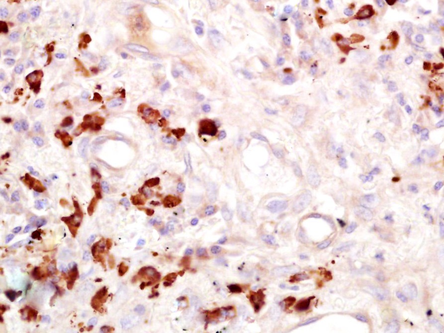 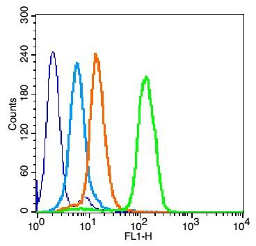 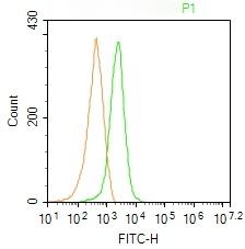 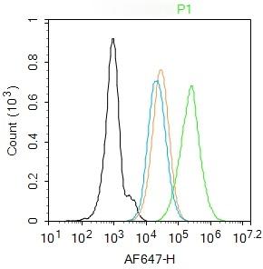 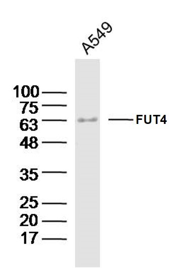 |
| 应用 | FCMIFIHC-FrIHC-PWB |
| 推荐剂量 | WB: 1:500-2000; IHC-P: 1:100-500; IHC-Fr: 1:100-500; IF: 1:100-500; FCM: 1μg/Test |
| 抗体种类 | Polyclonal |
| 宿主来源 | Rabbit |
| 亚细胞定位 | Golgi apparatus, Golgi stack membrane; Single-pass type II membrane protein. Note=Membrane-bound form in trans cisternae of Golgi. |
| 组织特异性 | Highest expression in stomach and colon. It is also expressed in the lung, testis, uterus, small intestine and to a lesser extent in spleen, and ovary. Present in trace amounts in brain, thymus, heart, smooth muscle, kidney and bone marrow. Not found in l |
| 构建方式 | Polyclonal Antibody |
| 纯化方式 | Protein A purified |
| 性状 | Liquid |
| 缓冲液 | 0.01M TBS (pH7.4) with 1% BSA, 0.02% Proclin300 and 50% Glycerol. |
| 浓度 | 1 mg/mL |
| 研究背景 | The Lewis histo-blood group system comprises a set of fucosylated glycosphingolipids that are synthesized by exocrine epithelial cells and circulate in body fluids. The glycosphingolipids function in embryogenesis, tissue differentiation, tumor metastasis, inflammation, and bacterial adhesion. They are secondarily absorbed to red blood cells giving rise to their Lewis phenotype. This gene is a member of the fucosyltransferase family, which catalyzes the addition of fucose to precursor polysaccharides in the last step of Lewis antigen biosynthesis. It encodes an enzyme with alpha(1,3)-fucosyltransferase and alpha(1,4)-fucosyltransferase activities. Mutations in this gene are responsible for the majority of Lewis antigen-negative phenotypes. Multiple alternatively spliced variants, encoding the same protein, have been found for this gene. [provided by RefSeq]. |
| 免疫原 | KLH conjugated synthetic peptide: human FUT4 |
| 抗原种属 | Human |
| 基因名称 | FUT4 |
| 基因ID | |
| 蛋白名称 | alpha-(1, 3)-fucosyltransferase 4 |
| Uniprot ID | |
| 研究领域 | CD markers,Surface molecules,Adhesion,Surface Molecules,Endothelial Markers,Human Lineage Negative,Tumor Associated,Golgi |
| 功能 | May catalyze alpha-1,3 glycosidic linkages involved in the expression of Lewis X/SSEA-1 and VIM-2 antigens. |
| 分子量 | Theoretical: 58 kDa. Actual: 63 kDa. |
| 储存方式 | Store at -20°C or -80°C for 12 months. Avoid repeated freeze-thaw cycles. |
| 运输方式 | Shipping with blue ice. |
 嗨!有任何问题?点我咨询
版权所有©2015-2026 TargetMol Chemicals Inc.保留所有权利.
沪ICP备20019793号-4 | 沪公网安备 31010602006700号 | 沪(静)应急管危经许[2024]203441