

购物车
 您的购物车当前为空

 很棒
很棒HOE 33187是一种DNA 染料,可渗透进入细胞。
很棒纯度: 99.86%
HOE 33187是一种DNA 染料,可渗透进入细胞。
| 规格 | 价格 | 库存 | 数量 |
|---|---|---|---|
| 5 mg | ¥ 163 | 现货 | |
| 10 mg | ¥ 239 | 现货 | |
| 25 mg | ¥ 392 | 现货 | |
| 50 mg | ¥ 648 | 现货 |
TargetMol的所有产品仅用作科学研究或药证申报,不能被用于人体,我们不向个人提供产品和服务。请您遵守承诺用途,不得违反法律法规规定用于任何其他用途。

| 产品描述 | Hoechst stains are part of a family of blue fluorescent dyes used to stain DNA. HOE 33187 is a cell dye for DNA. Hoechst dyes are cell-permeable and can bind to DNA in live or fixed cells. |
| 细胞实验 | 一、细胞核染色 1.材料准备: (1) Hoechst 33342溶液: 将Hoechst 33342溶解在适当的溶剂中,如水或PBS,常用浓度为1-10 µg/mL。 (2) 细胞样品: 活细胞或固定细胞,放在载玻片上或培养中。 (3) PBS缓冲液: 用于清洗细胞并调整pH。 2. 实验步骤: 活细胞染色 1)将Hoechst 33342加入细胞培养基中,达到推荐的最终浓度。 2)在室温或37°C下孵育15-30分钟,具体时间取决于细胞类型。 3)用PBS缓冲液清洗细胞,去除多余的染料。 4)使用荧光显微镜或流式细胞仪分析染色后的细胞。 激发波长:350 nm 发射波长:461 nm 固定细胞染色: 1)使用适当的固定液(如4%多聚甲醛)固定细胞。 2)固定后,按上述方法染色细胞,可以在载玻片上或悬浮液中染色。 3)使用显微镜分析细胞的荧光。 二、 细胞周期分析应用 1.按照上述方法染色凋亡细胞。 2.检查细胞的形态变化,如染色质凝缩和核碎片化,这些是凋亡的标志。 以上信息均来源于已发表文献,具体实验方案请根据实际研究需求进行适当调整。 |
| 分子量 | 408.5 |
| 分子式 | C25H24N6 |
| CAS No. | 23623-08-7 |
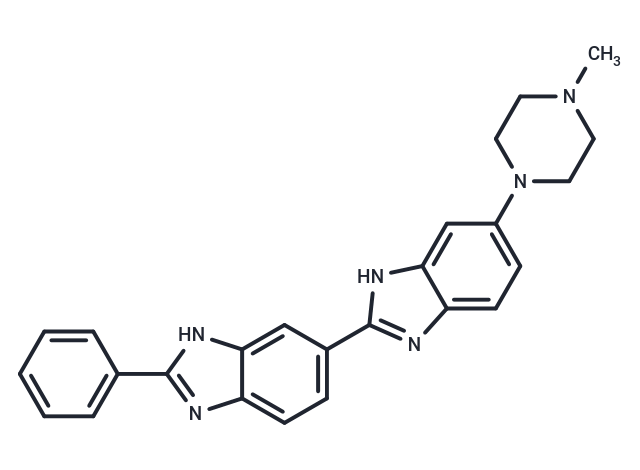
| Smiles | CN1CCN(CC1)c1ccc2nc([nH]c2c1)-c1ccc2nc([nH]c2c1)-c1ccccc1 |
| 密度 | 1.298 g/cm3 (Predicted) |
| 存储 | Keep away from direct sunlight Powder: -20°C for 3 years | In solvent: -80°C for 1 year Shipping with blue ice/Shipping at ambient temperature. 实际储存温度请以COA为准 | ||||||||||
| 溶解度信息 | DMSO: 2 mg/mL (4.9 mM), Sonication is recommended. | ||||||||||
溶液配制表 | |||||||||||
DMSO
该溶液配制表仅适用于固体产品。对于液体产品,请根据标明的浓度或密度计算稀释方案。 | |||||||||||
对于不同动物的给药剂量换算,您也可以参考 更多
 嗨!有任何问题?点我咨询
版权所有©2015-2026 TargetMol Chemicals Inc.保留所有权利.
沪ICP备20019793号-4 | 沪公网安备 31010602006700号 | 沪(静)应急管危经许[2024]203441