

购物车
 您的购物车当前为空


 还可以
还可以Anti-TMEM49 Polyclonal Antibody 是一种 Rabbit 抗体,靶向 TMEM49。Anti-TMEM49 Polyclonal Antibody 可用于 IF,IHC-Fr,IHC-P,WB。
还可以别名 VMP1, Vacuole membrane protein 1, Transmembrane protein 49, TMEM 49, TDC1, DKFZP566I133
Anti-TMEM49 Polyclonal Antibody 是一种 Rabbit 抗体,靶向 TMEM49。Anti-TMEM49 Polyclonal Antibody 可用于 IF,IHC-Fr,IHC-P,WB。
| 规格 | 价格 | 库存 | 数量 |
|---|---|---|---|
| 50 μL | ¥ 1,175 | 5日内发货 | |
| 100 μL | ¥ 1,965 | 5日内发货 | |
| 200 μL | ¥ 2,780 | 5日内发货 |
TargetMol的所有产品仅用作科学研究或药证申报,不能被用于人体,我们不向个人提供产品和服务。请您遵守承诺用途,不得违反法律法规规定用于任何其他用途。
| 产品描述 | Anti-TMEM49 Polyclonal Antibody is a Rabbit antibody targeting TMEM49. Anti-TMEM49 Polyclonal Antibody can be used in IF,IHC-Fr,IHC-P,WB. |
| 别名 | VMP1, Vacuole membrane protein 1, Transmembrane protein 49, TMEM 49, TDC1, DKFZP566I133 |
| Ig Type | IgG |
| 反应种属 | Mouse (predicted:Human,Rat,Pig,Horse,Rabbit) |
| 验证活性 | 1. Sample: Kidney (Mouse) Lysate at 40 μg Primary: Anti-TMEM49 (TMAB-01867) at 1/1000 dilution Secondary: IRDye800CW Goat Anti-Rabbit IgG at 1/20000 dilution Predicted band size: 46 kDa Observed band size: 46 kDa 2. Tissue/cell: mouse pancreas tissue; 4% Paraformaldehyde-fixed and paraffin-embedded; Antigen retrieval: citrate buffer (0.01M, pH6.0), Boiling bathing for 15 min; Block endogenous peroxidase by 3% Hydrogen peroxide for 30 min; Blocking buffer (normal goat serum) at 37°C for 20 min; Incubation: Anti-TMEM49 Polyclonal Antibody, Unconjugated (TMAB-01867) 1:200, overnight at 4°C, followed by conjugation to the secondary antibody and DAb staining. 3. Sample: Lane 1: Mouse Stomach tissue lysates Lane 2: Mouse Cerebrum tissue lysates Primary: Anti-TMEM49 (TMAB-01867) at 1/1000 dilution Secondary: IRDye800CW Goat Anti-Rabbit IgG at 1/20000 dilution Predicted band size: 46 kDa Observed band size: 48 kDa 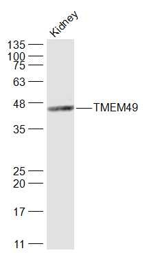 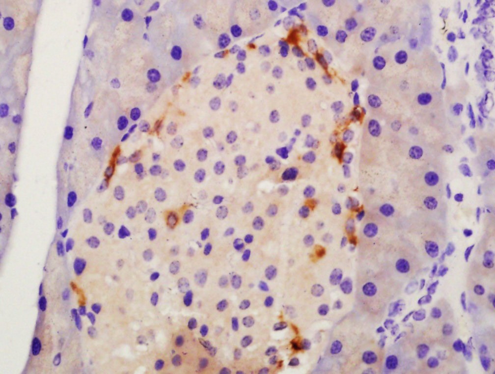 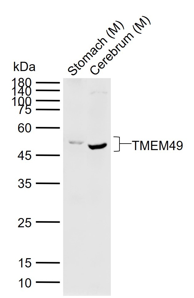 |
| 应用 | IFIHC-FrIHC-PWB |
| 推荐剂量 | WB: 1:500-2000; IHC-P: 1:100-500; IHC-Fr: 1:100-500; IF: 1:100-500 |
| 抗体种类 | Polyclonal |
| 宿主来源 | Rabbit |
| 亚细胞定位 | Endoplasmic reticulum-Golgi intermediate compartment membrane. Cell membrane. Vacuole membrane. Endoplasmic reticulum. |
| 构建方式 | Polyclonal Antibody |
| 纯化方式 | Protein A purified |
| 性状 | Liquid |
| 缓冲液 | 0.01M TBS (pH7.4) with 1% BSA, 0.02% Proclin300 and 50% Glycerol. |
| 浓度 | 1 mg/mL |
| 研究背景 | Vacuole membrane protein 1 (VMP1)/TMEM49 is a transmembrane protein localized to intracellular vacuoles and was discovered as a protein that promotes vacuole formation in acinar cells associated with acute pancreatitis (1). Over-expression of VMP1 promotes vacuole formation and subsequent cell death (1). Subsequent studies have shown that VMP1 expression is induced by starvation and the mTOR inhibitor, rapamycin, and can trigger autophagy (2). VMP1 is targeted, along with LC3, to autophagosome membranes (2). Knockdown of VMP1 can inhibit autophagosome formation (2). VMP1 interacts with Beclin-1, a key autophagy protein that activates the Class III PI3 kinase Vps34, which is regulated by a large network of associated proteins (3). VMP1 functions in the degradation and clearance of zymogen-containing vacuoles during experimental pancreatitis (4). During this process, VMP1 interacts with the ubiquitin protease USP9X, suggesting a possible functional link between the molecular machinery of autophagy and the ubiquitin pathway. Orthologues of VMP1 have been reported in C. elegans (known as EPG-3), Drosophila (known as TANGO-5), and Dictyostelium, and have been shown to play a role in membrane trafficking, orgenelle organization, and autophagy (5-7). |
| 免疫原 | KLH conjugated synthetic peptide: human TMEM49 |
| 抗原种属 | Human |
| 基因名称 | VMP1 |
| 基因ID | |
| 蛋白名称 | Vacuole membrane protein 1 |
| Uniprot ID | |
| 研究领域 | Autophagosome,Tumor biomarkers,Autophagosome,Autophagosome |
| 功能 | Stress-induced protein that, when overexpressed, promotes formation of intracellular vacuoles followed by cell death. May be involved in the cytoplasmic vacuolization of acinar cells during the early stage of acute pancreatitis. Plays a role in the initial stages of the autophagic process through its interaction with BECN1 (By similarity). Involved in cell-cell adhesion. Plays an essential role in formation of cell junctions.Sequence similarities: Belongs to the VMP1 family. |
| 分子量 | Theoretical: 46 kDa. Actual: 48 kDa. |
| 储存方式 | Store at -20°C or -80°C for 12 months. Avoid repeated freeze-thaw cycles. |
| 运输方式 | Shipping with blue ice. |
 嗨!有任何问题?点我咨询
版权所有©2015-2026 TargetMol Chemicals Inc.保留所有权利.
沪ICP备20019793号-4 | 沪公网安备 31010602006700号 | 沪(静)应急管危经许[2024]203441