

购物车
 您的购物车当前为空


 还可以
还可以Anti-HK1 Antibody (7G677) 是一种 Rabbit 抗体,靶向 HK1。Anti-HK1 Antibody (7G677) 可用于 FCM,ICC/IF,IHC,IP,WB。
还可以别名 HK1, Hexokinase-A, Hexokinase-1, Hexokinase type I (HK I), Brain form hexokinase
Anti-HK1 Antibody (7G677) 是一种 Rabbit 抗体,靶向 HK1。Anti-HK1 Antibody (7G677) 可用于 FCM,ICC/IF,IHC,IP,WB。
| 规格 | 价格 | 库存 | 数量 |
|---|---|---|---|
| 50 μL | ¥ 1,490 | 5日内发货 | |
| 100 μL | ¥ 2,495 | 5日内发货 |
TargetMol的所有产品仅用作科学研究或药证申报,不能被用于人体,我们不向个人提供产品和服务。请您遵守承诺用途,不得违反法律法规规定用于任何其他用途。
| 产品描述 | Anti-HK1 Antibody (7G677) is a Rabbit antibody targeting HK1. Anti-HK1 Antibody (7G677) can be used in FCM,ICC/IF,IHC,IP,WB. |
| 别名 | HK1, Hexokinase-A, Hexokinase-1, Hexokinase type I (HK I), Brain form hexokinase |
| Ig Type | IgG |
| 克隆号 | 7G677 |
| 反应种属 | Human,Mouse,Rat |
| 验证活性 | 1. Western blot analysis of Hexokinase 1 on different lysates using anti-Hexokinase 1 antibody at 1/1,000 dilution. Positive control: Lane 1: Hela, Lane 2: 293, Lane 3: MCF-7, Lane 4: HepG2. 2. Immunohistochemical analysis of paraffin-embedded mouse skin tissue using anti-Hexokinase 1 antibody. Counter stained with hematoxylin. 3. Immunohistochemical analysis of paraffin-embedded human lung tissue using anti-Hexokinase 1 antibody. Counter stained with hematoxylin. 4. Immunohistochemical analysis of paraffin-embedded human breast carcinoma tissue using anti-Hexokinase 1 antibody. Counter stained with hematoxylin. 5. Immunohistochemical analysis of paraffin-embedded human kidney tissue using anti-Hexokinase 1 antibody. Counter stained with hematoxylin. 6. Immunohistochemical analysis of paraffin-embedded mouse colon tissue using anti-Hexokinase 1 antibody. Counter stained with hematoxylin. 7. Immunohistochemical analysis of paraffin-embedded mouse brain tissue using anti-Hexokinase 1 antibody. Counter stained with hematoxylin. 8. ICC staining Hexokinase 1 in Hela cells (green). The nuclear counter stain is DAPI (blue). Cells were fixed in paraformaldehyde, permeabilised with 0.25% Triton X100/PBS. 9. ICC staining Hexokinase 1 in CRC cells (green). The nuclear counter stain is DAPI (blue). Cells were fixed in paraformaldehyde, permeabilised with 0.25% Triton X100/PBS. 10. ICC staining Hexokinase 1 in MCF-7 cells (green). The nuclear counter stain is DAPI (blue). Cells were fixed in paraformaldehyde, permeabilised with 0.25% Triton X100/PBS. 11. Flow cytometric analysis of K562 cells with Hexokinase 1 antibody at 1/50 dilution (blue) compared with an unlabelled control (cells without incubation with primary antibody; red). Alexa Fluor 488-conjugated goat anti rabbit IgG was used as the secondary antibody. 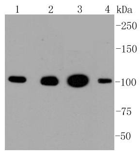 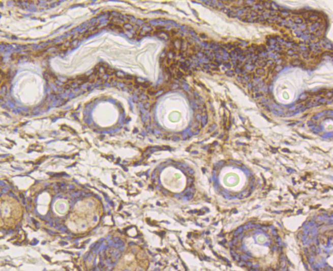 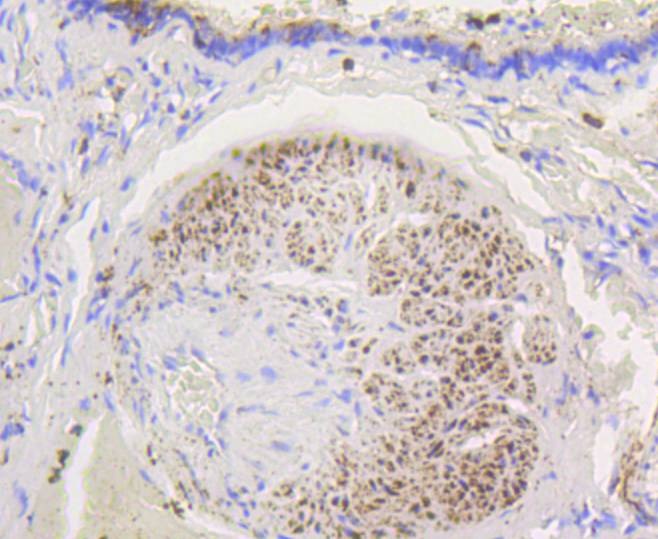 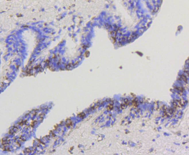 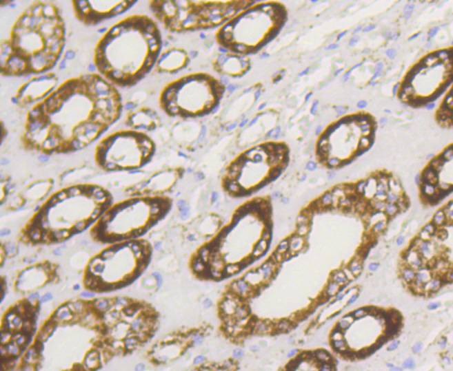 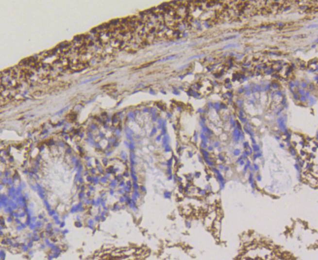 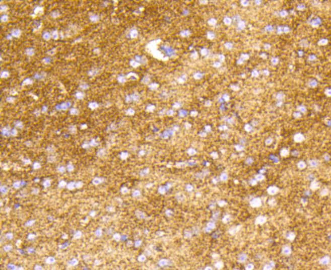 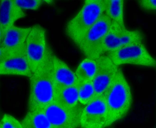 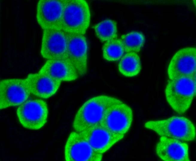 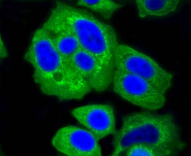 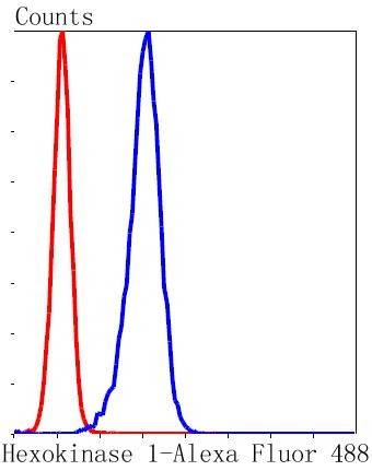 |
| 应用 | FCMICC/IFIHCIPWB |
| 推荐剂量 | WB: 1:1000-2000; IHC: 1:50-200; ICC/IF: 1:50-200; FCM: 1:50-100 |
| 抗体种类 | Monoclonal |
| 宿主来源 | Rabbit |
| 构建方式 | Recombinant Antibody |
| 纯化方式 | ProA affinity purified |
| 性状 | Liquid |
| 缓冲液 | 1*TBS (pH7.4), 1%BSA, 40%Glycerol. Preservative: 0.05% Sodium Azide. |
| 研究背景 | The hexokinases utilize Mg-ATP as a phosphoryl donor to catalyze the first step of intracellular glucose metabolism, the conversion of glucose to glucose-6-phosphate. Four hexokinase isoenzymes have been identified, including hexokinase I (HXK I), hexokinase II (HXK II), hexokinase III (HXK III) and hexokinase IV (HXK IV, also designated glucokinase or GCK). Hexokinases I-III each contain an N-terminal cluster of hydrophobic amino acids. Glucokinase lacks the N-terminal hydrophobic cluster. The hydrophobic cluster is thought to be necessary for membrane binding. This is substantiated by the finding that glucokinase has lower affinity for glucose than do the other hexokinases. HXK I has been shown to be expressed in brain, kidney and heart tissues as well as in hepatoma cell lines. HXK II is involved in the uptake and utilization of glucose by adipose and skeletal tissues. Of the hexokinases, HXK III has the highest affinity for glucose. Glucokinase is expressed in pancreatic beta cells where it functions as a glucose sensor, determining the ??set point?? for insulin secretion. |
| 偶联 | Unconjugated |
| 免疫原 | Recombinant Protein |
| Uniprot ID |
| 分子量 | Theoretical: 102 kDa. |
| 储存方式 | Store at -20°C or -80°C for 12 months. Avoid repeated freeze-thaw cycles. |
| 运输方式 | Shipping with blue ice. |
 嗨!有任何问题?点我咨询
版权所有©2015-2026 TargetMol Chemicals Inc.保留所有权利.
沪ICP备20019793号-4 | 沪公网安备 31010602006700号 | 沪(静)应急管危经许[2024]203441