

购物车
 您的购物车当前为空


 还可以
还可以Anti-SIRT1 Polyclonal Antibody 是一种 Rabbit 抗体,靶向 SIRT1。Anti-SIRT1 Polyclonal Antibody 可用于 FCM,IF,IHC-Fr,IHC-P,WB。
还可以别名 sirtuin 1, SIR2L1
Anti-SIRT1 Polyclonal Antibody 是一种 Rabbit 抗体,靶向 SIRT1。Anti-SIRT1 Polyclonal Antibody 可用于 FCM,IF,IHC-Fr,IHC-P,WB。
| 规格 | 价格 | 库存 | 数量 |
|---|---|---|---|
| 50 μL | ¥ 1,160 | 5日内发货 | |
| 100 μL | ¥ 1,960 | 5日内发货 | |
| 200 μL | ¥ 2,780 | 5日内发货 |
TargetMol的所有产品仅用作科学研究或药证申报,不能被用于人体,我们不向个人提供产品和服务。请您遵守承诺用途,不得违反法律法规规定用于任何其他用途。
| 产品描述 | Anti-SIRT1 Polyclonal Antibody is a Rabbit antibody targeting SIRT1. Anti-SIRT1 Polyclonal Antibody can be used in FCM,IF,IHC-Fr,IHC-P,WB. |
| 别名 | sirtuin 1, SIR2L1 |
| Ig Type | IgG |
| 反应种属 | Human,Mouse (predicted:Dog,Pig,Horse,Rabbit) |
| 验证活性 | 1. Blank control (blue line): MCF 7 (blue). Primary Antibody (green line): Rabbit Anti-SIRT1 antibody (TMAB-01690) Dilution: 3 μg/10^5 cells; Isotype Control Antibody (orange line): Rabbit IgG. Secondary Antibody (white blue line): Goat anti-rabbit IgG-FITC Dilution: 1 μg/test. Protocol The cells were fixed with 70% methanol (Overnight at 4°C) and then permeabilized with 90% ice-cold methanol for 30 min on ice. Cells stained with Primary Antibody for 30 min at room temperature. The cells were then incubated in 1 X PBS/2% BSA/10% goat serum to block non-specific protein-protein interactions followed by the antibody for 15 min at room temperature. The secondary antibody used for 40 min at room temperature. 2. Blank control: HL-60. Primary Antibody (green line): Rabbit Anti-SIRT1 antibody (TMAB-01690) Dilution: 1 μg/10^6 cells; Isotype Control Antibody (orange line): Rabbit IgG. Secondary Antibody: Goat anti-rabbit IgG-AF488 Dilution: 1 μg/test. Protocol The cells were fixed with 4% PFA (10 min at room temperature) and then permeabilized with 0.1% PBST for 20 min at room temperature. The cells were then incubated in 5% BSA to block non-specific protein-protein interactions for 30 min at room temperature. Cells stained with Primary Antibody for 30 min at room temperature. The secondary antibody used for 40 min at room temperature. 3. Sample: Lane 1: Hela (Human) Cell Lysate at 30 μg Lane 2: NIH/3T3 (Mouse) Cell Lysate at 30 μg Lane 3: HepG2 (Human) Cell Lysate at 30 μg Lane 4: 293T (Human) Cell Lysate at 30 μg Lane 5: Testis (Mouse) Lysate at 40 μg Primary: Anti-SIRT1 (TMAB-01690) at 1/1000 dilution Secondary: IRDye800CW Goat Anti-Rabbit IgG at 1/20000 dilution Predicted band size: 116/95 kDa Observed band size: 116 kDa 4. Paraformaldehyde-fixed, paraffin embedded (human glioma tissue); Antigen retrieval by boiling in sodium citrate buffer (pH6.0) for 15 min; Block endogenous peroxidase by 3% hydrogen peroxide for 20 min; Blocking buffer (normal goat serum) at 37°C for 30 min; Antibody incubation with (SIRT1) Polyclonal Antibody, Unconjugated (TMAB-01690) at 1:200 overnight at 4°C, followed by operating according to SP Kit (Rabbit) instructionsand DAB staining. 5. Blank control: HL-60. Primary Antibody (green line): Rabbit Anti-SIRT1 antibody Dilution: 1 μg/10^6 cells; Isotype Control Antibody (orange line): Rabbit IgG. Secondary Antibody: Goat anti-rabbit IgG-AF488 Dilution: 1 μg/test. Protocol The cells were fixed with 4% PFA (10 min at room temperature) and then permeabilized with 0.1% PBST for 20 min at room temperature. The cells were then incubated in 5% BSA to block non-specific protein-protein interactions for 30 min at room temperature. Cells stained with Primary Antibody for 30 min at room temperature. The secondary antibody used for 40 min at room temperature. 6. Paraformaldehyde-fixed, paraffin embedded (mouse brain); Antigen retrieval by boiling in sodium citrate buffer (pH6.0) for 15 min; Block endogenous peroxidase by 3% hydrogen peroxide for 20 min; Blocking buffer (normal goat serum) at 37°C for 30 min; Antibody incubation with (SIRT1) Polyclonal Antibody, Unconjugated (TMAB-01690) at 1:200 overnight at 4°C, followed by operating according to SP Kit (Rabbit) instructionsand DAB staining. 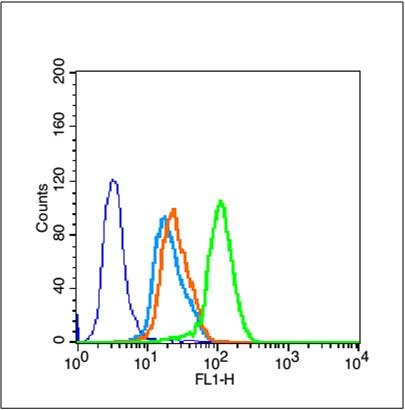 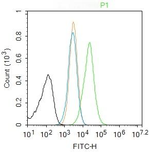 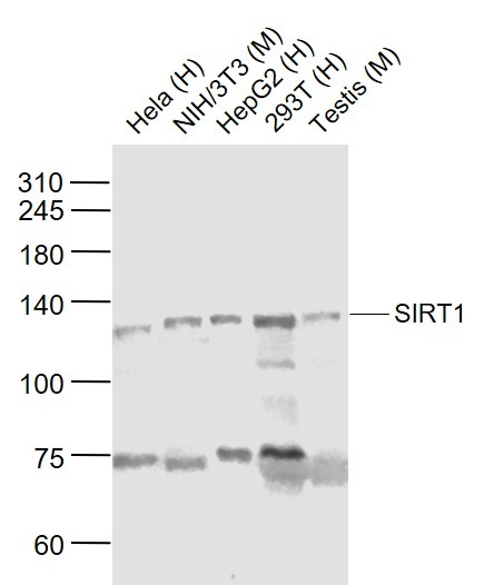 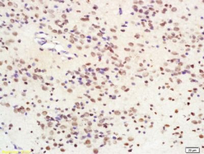 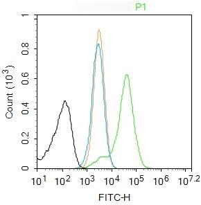 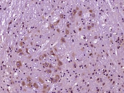 |
| 应用 | FCMIFIHC-FrIHC-PWB |
| 推荐剂量 | WB: 1:500-2000; IHC-P: 1:100-500; IHC-Fr: 1:100-500; IF: 1:100-500; FCM: 1ug/Test |
| 抗体种类 | Polyclonal |
| 宿主来源 | Rabbit |
| 亚细胞定位 | Nucleus, PML body. Cytoplasm. Note=Recruited to the nuclear bodies via its interaction with PML. Colocalized with APEX1 in the nucleus. May be found in nucleolus, nuclear euchromatin, heterochromatin and inner membrane. Shuttles between nucleus and cytoplasm.SirtT1 75 kDa fragment: Cytoplasm. Mitochondrion. |
| 组织特异性 | Widely expressed. |
| 构建方式 | Polyclonal Antibody |
| 纯化方式 | Protein A purified |
| 性状 | Liquid |
| 缓冲液 | 0.01M TBS (pH7.4) with 1% BSA, 0.02% Proclin300 and 50% Glycerol. |
| 浓度 | 1 mg/mL |
| 研究背景 | This gene encodes a member of the sirtuin family of proteins, homologs to the yeast Sir2 protein. Members of the sirtuin family are characterized by a sirtuin core domain and grouped into four classes. The functions of human sirtuins have not yet been determined; however, yeast sirtuin proteins are known to regulate epigenetic gene silencing and suppress recombination of rDNA. Studies suggest that the human sirtuins may function as intracellular regulatory proteins with mono-ADP-ribosyltransferase activity. The protein encoded by this gene is included in class I of the sirtuin family. Alternative splicing results in multiple transcript variants. Function : SirtT1 75 kDa fragment: catalytically inactive 75SirT1 may be involved in regulation of apoptosis. May be involved in protecting chondrocytes from apoptotic death by associating with cytochrome C and interfering with apoptosome assembly. Subunit : Found in a complex with PCAF and MYOD1. Component of the eNoSC complex, composed of SIRT1, SUV39H1 and RRP8. Interacts with HES1, HEY2 and PML. Interacts with RPS19BP1/AROS. Interacts with KIAA1967/DBC1 (via N-terminus); the interaction disrupts the interaction between SIRT1 and p53/TP53. Interacts with SETD7; the interaction induces the dissociation of SIRT1 from p53/TP53 and increases p53/TP53 activity. Interacts with MYCN, NR1I2, CREBZF, TSC2, TLE1, FOS, JUN, NR0B2, PPARG, NCOR, IRS1, IRS2 and NMNAT1. Interacts with HNF1A; the interaction occurs under nutrient restriction. Interacts with SUZ12; the interaction mediates the association with the PRC4 histone methylation complex which is specific as an association with PCR2 and PCR3 complex variants is not found. Interacts with HIV-1 tat. Subcellular Location : Nucleus, PML body. Cytoplasm. Note=Recruited to the nuclear bodies via its interaction with PML. Colocalized with APEX1 in the nucleus. May be found in nucleolus, nuclear euchromatin, heterochromatin and inner membrane. Shuttles between nucleus and cytoplasm. SirtT1 75 kDa fragment: Cytoplasm. Mitochondrion. Tissue Specificity : Widely expressed. Post-translational modifications : Methylated on multiple lysine residues; methylation is enhanced after DNA damage and is dispensable for deacetylase activity toward p53/TP53. Phosphorylated. Phosphorylated by STK4/MST1, resulting in inhibition of SIRT1-mediated p53/TP53 deacetylation. Phosphorylation by MAPK8/JNK1 at Ser-27, Ser-47, and Thr-530 leads to increased nuclear localization and enzymatic activity. Phosphorylation at Thr-530 by DYRK1A and DYRK3 acivates deacetylase activity and promotes cell survival. Phosphorylation by mammalian target of rapamycin complex 1 (mTORC1) at Ser-47 inhibits deacetylation activity. Phosphorylated by CaMK2, leading to increased p53/TP53 and NF-kappa-B p65/RELA deacetylation activity (By similarity). Phosphorylation at Ser-27 implicating MAPK9 is linked to protein stability. There is some ambiguity for some phosphosites: Ser-159/Ser-162 and Thr-544/Ser-545. Proteolytically cleaved by cathepsin B upon TNF-alpha treatment to yield catalytic inactive but stable SirtT1 75 kDa fragment (75SirT1). S-nitrosylated by GAPDH, leading to inhibit the NAD-dependent protein deacetylase activity (By similarity). Similarity : Belongs to the sirtuin family. Contains 1 deacetylase sirtuin-type domain. |
| 免疫原 | KLH conjugated synthetic peptide: human SirtT1 |
| 抗原种属 | Human |
| 基因名称 | SIRT1 |
| 基因ID | |
| 蛋白名称 | NAD-dependent protein deacetylase sirtuin-1 |
| Uniprot ID | |
| 研究领域 | p53 Pathway,Acetylation,Class III / Sir2 class,Obesity,Host Virus Interaction,Other Nuclear Bodies |
| 功能 | SirtT1 75 kDa fragment: catalytically inactive 75SirT1 may be involved in regulation of apoptosis. May be involved in protecting chondrocytes from apoptotic death by associating with cytochrome C and interfering with apoptosome assembly. |
| 分子量 | Theoretical: 58/81 kDa. Actual: 116 kDa. |
| 储存方式 | Store at -20°C or -80°C for 12 months. Avoid repeated freeze-thaw cycles. |
| 运输方式 | Shipping with blue ice. |
 嗨!有任何问题?点我咨询
版权所有©2015-2026 TargetMol Chemicals Inc.保留所有权利.
沪ICP备20019793号-4 | 沪公网安备 31010602006700号 | 沪(静)应急管危经许[2024]203441