

购物车
 您的购物车当前为空


 还可以
还可以Anti-Pan Cytokeratin Polyclonal Antibody 2 是一种 Rabbit 抗体,靶向 Pan Cytokeratin。Anti-Pan Cytokeratin Polyclonal Antibody 2 可用于 FCM, ICC/IF, IF, IHC-Fr, IHC-P, WB。
还可以别名 wide spectrum Cytokeratin, P-CK, pan-cytokeratin, pan-CK, Cytokeratins, CK-PAN, [cytokeratins 13, 14, 16, 17, 19, 24]
Anti-Pan Cytokeratin Polyclonal Antibody 2 是一种 Rabbit 抗体,靶向 Pan Cytokeratin。Anti-Pan Cytokeratin Polyclonal Antibody 2 可用于 FCM, ICC/IF, IF, IHC-Fr, IHC-P, WB。
| 规格 | 价格 | 库存 | 数量 |
|---|---|---|---|
| 50 μL | ¥ 1,175 | 5日内发货 | |
| 100 μL | ¥ 1,960 | 5日内发货 | |
| 200 μL | ¥ 2,790 | 5日内发货 |
TargetMol的所有产品仅用作科学研究或药证申报,不能被用于人体,我们不向个人提供产品和服务。请您遵守承诺用途,不得违反法律法规规定用于任何其他用途。
| 产品描述 | Anti-Pan Cytokeratin Polyclonal Antibody 2 is a Rabbit antibody targeting Pan Cytokeratin. Anti-Pan Cytokeratin Polyclonal Antibody 2 can be used in FCM, ICC/IF, IF, IHC-Fr, IHC-P, WB. |
| 别名 | wide spectrum Cytokeratin, P-CK, pan-cytokeratin, pan-CK, Cytokeratins, CK-PAN, [cytokeratins 13, 14, 16, 17, 19, 24] |
| Ig Type | IgG |
| 反应种属 | Human,Mouse,Rat (predicted:Chicken,Dog,Pig,Cow,Horse,Rabbit) |
| 验证活性 | 1. Paraformaldehyde-fixed, paraffin embedded (Rat bladder); Antigen retrieval by boiling in sodium citrate buffer (pH6.0) for 15 min; Block endogenous peroxidase by 3% hydrogen peroxide for 20 min; Blocking buffer (normal goat serum) at 37°C for 30 min; Antibody incubation with (Pan Cytokeratin) Polyclonal Antibody, Unconjugated (TMAB-01330) at 1:400 overnight at 4°C, followed by operating according to SP Kit (Rabbit) instructionsand DAB staining. 2. Sample: Bladder (Mouse) Lysate at 40 μg Primary: Anti-Pan Cytokeratin (TMAB-01330) at 1/300 dilution Secondary: IRDye800CW Goat Anti-Rabbit IgG at 1/20000 dilution Predicted band size: 42-64 kDa Observed band size: 60 kDa 3. Blank control (blue line): Hela (blue). Primary Antibody (green line): Rabbit Anti-Pan Cytokeratin antibody (TMAB-01330) Dilution: 1 μg/10^6 cells; Isotype Control Antibody (orange line): Rabbit IgG. Secondary Antibody (white blue line): Goat anti-rabbit IgG-PE Dilution: 1 μg/test. Protocol The cells were fixed with 70% methanol (Overnight at 4°C) and then permeabilized with 90% ice-cold methanol for 20 min at-20°C. Cells stained with Primary Antibody for 30 min at room temperature. The cells were then incubated in 1 X PBS/2% BSA/10% goat serum to block non-specific protein-protein interactions followed by the antibody for 15 min at room temperature. The secondary antibody used for 40 min at room temperature. 4. Paraformaldehyde-fixed, paraffin embedded (Human stomach carcinoma); Antigen retrieval by boiling in sodium citrate buffer (pH6.0) for 15 min; Block endogenous peroxidase by 3% hydrogen peroxide for 20 min; Blocking buffer (normal goat serum) at 37°C for 30 min; Antibody incubation with (Pan Cytokeratin) Polyclonal Antibody, Unconjugated (TMAB-01330) at 1:400 overnight at 4°C, followed by operating according to SP Kit (Rabbit) instructionsand DAB staining. 5. Paraformaldehyde-fixed, paraffin embedded (human liver); Antigen retrieval by boiling in sodium citrate buffer (pH6.0) for 15 min; Block endogenous peroxidase by 3% hydrogen peroxide for 20 min; Blocking buffer (normal goat serum) at 37°C for 30 min; Antibody incubation with (Pan Cytokeratin) Polyclonal Antibody, Unconjugated (TMAB-01330) at 1:200 overnight at 4°C, followed by operating according to SP Kit (Rabbit) instructionsand DAB staining. 6. Paraformaldehyde-fixed, paraffin embedded (Human kidney); Antigen retrieval by boiling in sodium citrate buffer (pH6.0) for 15 min; Block endogenous peroxidase by 3% hydrogen peroxide for 20 min; Blocking buffer (normal goat serum) at 37°C for 30 min; Antibody incubation with (Pan Cytokeratin) Polyclonal Antibody, Unconjugated (TMAB-01330) at 1:200 overnight at 4°C, followed by operating according to SP Kit (Rabbit) instructionsand DAB staining. 7. Sample: Lane 1: Hela (Human) Cell Lysate at 30 μg Lane 2: A549 (Human) Cell Lysate at 30 μg Lane 3: A673 (Human) Cell Lysate at 30 μg Primary: Anti-Pan Cytokeratin (TMAB-01330) at 1/1000 dilution Secondary: IRDye800CW Goat Anti-Rabbit IgG at 1/20000 dilution Predicted band size: 42-64 kDa Observed band size: 46,60 kDa 8. Paraformaldehyde-fixed, paraffin embedded (human cervical cancer); Antigen retrieval by boiling in sodium citrate buffer (pH6.0) for 15 min; Block endogenous peroxidase by 3% hydrogen peroxide for 20 min; Blocking buffer (normal goat serum) at 37°C for 30 min; Antibody incubation with (Pan Cytokeratin) Polyclonal Antibody, Unconjugated (TMAB-01330) at 1:200 overnight at 4°C, followed by operating according to SP Kit (Rabbit) instructionsand DAB staining. 9. Paraformaldehyde-fixed, paraffin embedded (rat uterus); Antigen retrieval by boiling in sodium citrate buffer (pH6.0) for 15 min; Block endogenous peroxidase by 3% hydrogen peroxide for 20 min; Blocking buffer (normal goat serum) at 37°C for 30 min; Antibody incubation with (Pan Cytokeratin) Polyclonal Antibody, Unconjugated (TMAB-01330) at 1:200 overnight at 4°C, followed by operating according to SP Kit (Rabbit) instructionsand DAB staining. 10. Hela cell; 4% Paraformaldehyde-fixed; Triton X-100 at room temperature for 20 min; Blocking buffer (normal goat serum) at 37°C for 20 min; Antibody incubation with (Pan Cytokeratin) polyclonal Antibody, Unconjugated (TMAB-01330) 1:100, 90 minutes at 37°C; followed by a conjugated Goat Anti-Rabbit IgG antibody at 37°C for 90 minutes, DAPI (blue) was used to stain the cell nucleus. 11. Paraformaldehyde-fixed, paraffin embedded (human breast carcinoma); Antigen retrieval by boiling in sodium citrate buffer (pH6.0) for 15 min; Block endogenous peroxidase by 3% hydrogen peroxide for 20 min; Blocking buffer (normal goat serum) at 37°C for 30 min; Antibody incubation with (Pan Cytokeratin) Polyclonal Antibody, Unconjugated (TMAB-01330) at 1:200 overnight at 4°C, followed by operating according to SP Kit (Rabbit) instructionsand DAB staining. 12. Paraformaldehyde-fixed, paraffin embedded (human colon carcinoma); Antigen retrieval by boiling in sodium citrate buffer (pH6.0) for 15 min; Blocking buffer (normal goat serum) at 37°C for 30 min; Antibody incubation with (Pan Cytokeratin) Polyclonal Antibody, Unconjugated (TMAB-01330) at 1:200 overnight at 4°C, followed by a conjugated Goat Anti-Rabbit IgG antibody for 90 minutes, and DAPI for nucleus staining. 13. Paraformaldehyde-fixed, paraffin embedded (human cervical carcinoma); Antigen retrieval by boiling in sodium citrate buffer (pH6.0) for 15 min; Block endogenous peroxidase by 3% hydrogen peroxide for 20 min; Blocking buffer (normal goat serum) at 37°C for 30 min; Antibody incubation with (Pan Cytokeratin) Polyclonal Antibody, Unconjugated (TMAB-01330) at 1:200 overnight at 4°C, followed by operating according to SP Kit (Rabbit) instructionsand DAB staining. 14. Blank control: Hela. Primary Antibody (green line): Rabbit Anti-Pan Cytokeratin antibody (TMAB-01330) Dilution: 2 μg/Test; Secondary Antibody: Goat anti-rabbit IgG-FITC Dilution: 0.5 μg/Test. Protocol The cells were fixed with 4% PFA (10 min at room temperature) and then permeabilized with 0.1% PBST for 20 min at room temperature. The cells were then incubated in 5% BSA to block non-specific protein-protein interactions for 30 min at room temperature. Cells stained with Primary Antibody for 30 min at room temperature. The secondary antibody used for 40 min at room temperature. 15. Blank control (black line): A549. Primary Antibody (green line): Rabbit Anti-Pan Cytokeratin antibody (TMAB-01330) Dilution: 1 μg/Test; Secondary Antibody (white blue line): Goat anti-rabbit IgG-AF488 Dilution: 0.5 μg/Test. Isotype control (orange line): Normal Rabbit IgG Protocol The cells were fixed with 4% PFA (10 min at room temperature) and then permeabilized with 90% ice-cold methanol for 20 min at-20°C, The cells were then incubated in 5% BSA to block non-specific protein-protein interactions for 30 min at room temperature. Cells stained with Primary Antibody for 30 min at room temperature. The secondary antibody used for 40 min at room temperature. 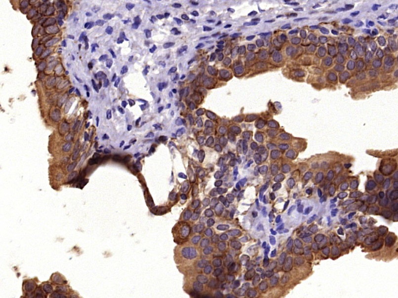 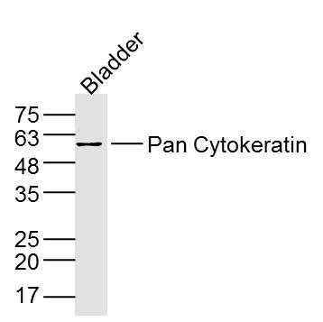 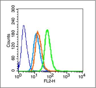 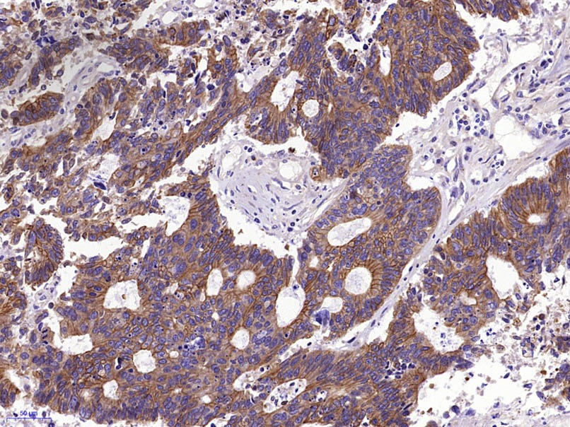 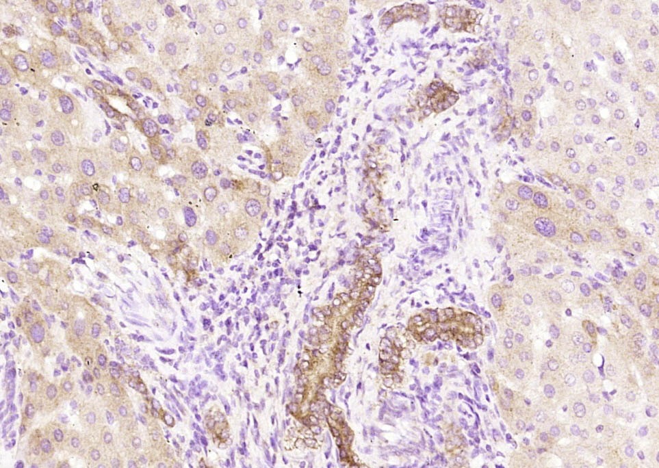 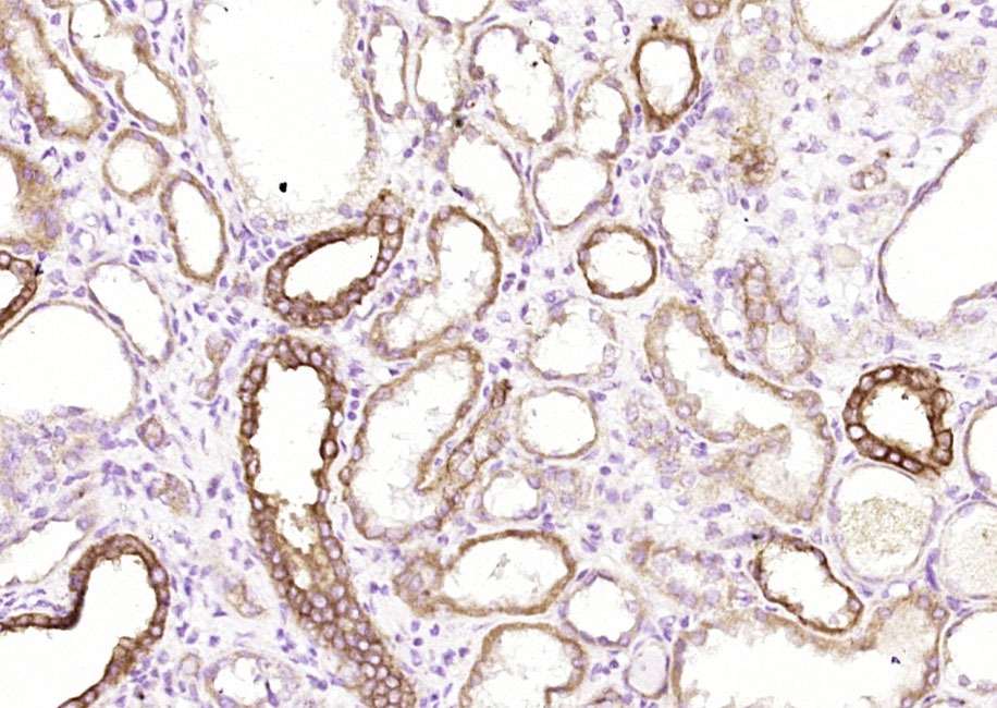 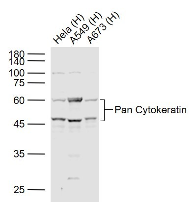 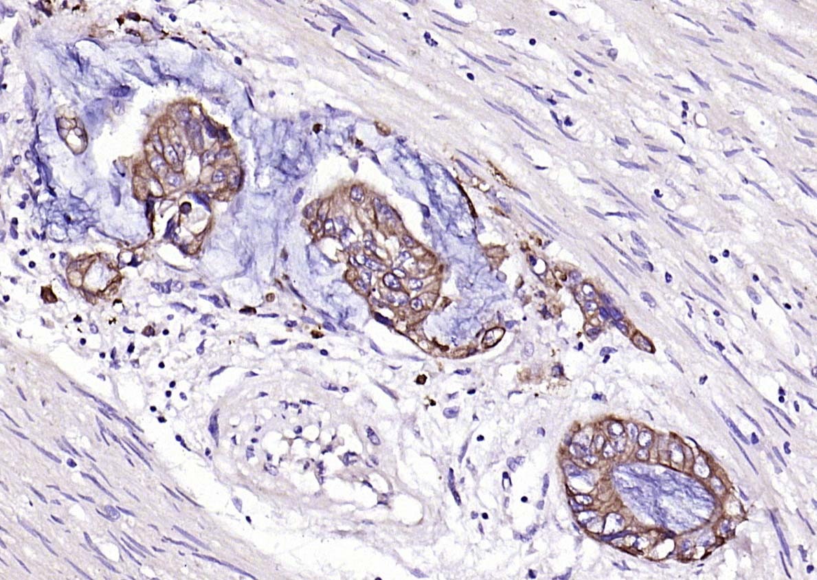 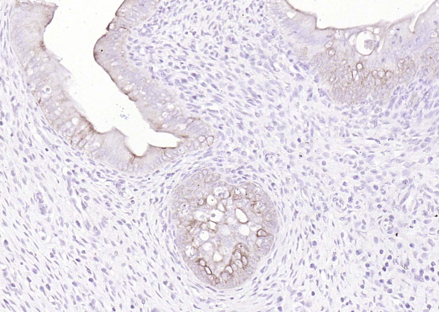 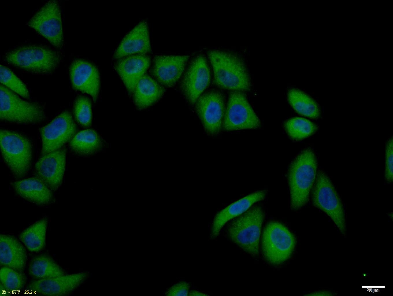  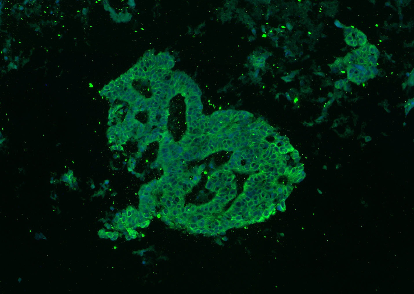 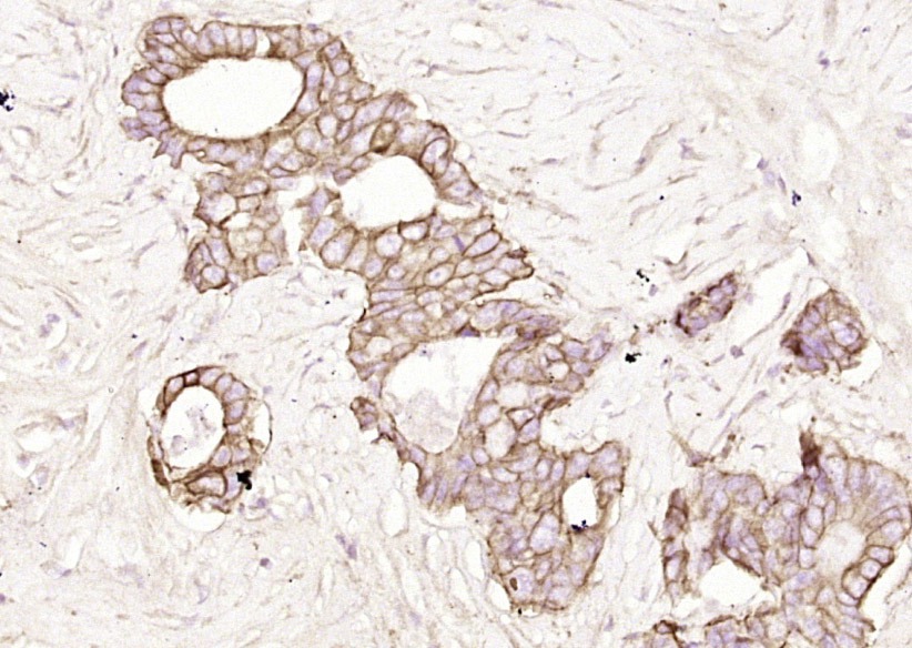 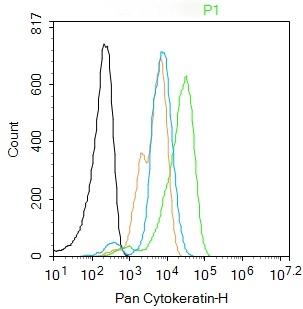 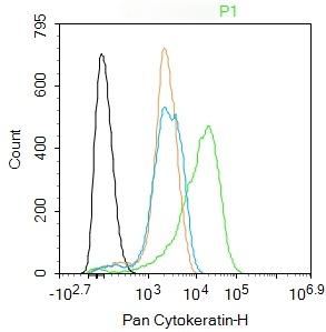 |
| 应用 | FCMICC/IFIFIHC-FrIHC-PWB |
| 推荐剂量 | FCM=1 μg/Test; ICC/IF=1:100-500; IF=1:100-500; IHC-Fr=1:100-500; IHC-P=1:100-500; WB=1:500-2000 |
| 抗体种类 | Polyclonal |
| 宿主来源 | Rabbit |
| 亚细胞定位 | Cytoplasmic. |
| 组织特异性 | epithelial cells |
| 构建方式 | Polyclonal Antibody |
| 纯化方式 | Protein A purified |
| 性状 | Liquid |
| 缓冲液 | 0.01M TBS (pH7.4) with 1% BSA, 0.02% Proclin300 and 50% Glycerol. |
| 浓度 | 1 mg/mL |
| 研究背景 | Cytokeratins are proteins of keratin-containing intermediate filaments found in the intracytoplasmic cytoskeleton of epithelial tissue. The cytokeratins are encoded by a family encompassing 30 genes. Among them, 20 are epithelial genes and the remaining 10 are specific for trichocytes. In the cytoplasm, the keratin filaments conform a complex network which extends from the surface of the nucleus to the cell membrane. Numerous accessory proteins are involved in the genesis and maintenance of such structure. This association between the plasma membrane and the nuclear surface provides important implications for the organization of the cytoplasm and cellular communication mechanisms. Apart from the relatively static functions provided in terms of supporting the nucleus and providing tensile strength to the cell, the cytokeratin networks undergo rapid phosphate exchanges mediated depolymerization, with important implications in the more dynamic cellular processes such as mitosis and post-mitotic period, cell movement and differentiation. Cytokeratins interact with desmosomes and hemidesmosomes, thus collaborating to cell-cell adhesion and basal cell-underlying connective tissue connection. |
| 免疫原 | KLH conjugated synthetic peptide: human Cytokeratin-1 |
| 抗原种属 | Human |
| 基因名称 | KRT24 |
| 基因ID | |
| 蛋白名称 | Keratin, type I cytoskeletal 24 |
| Uniprot ID | |
| 研究领域 | Cytokeratins |
| 分子量 | Theoretical: 42-64 kDa. Actual: 46,60 kDa. |
| 储存方式 | Store at -20°C or -80°C for 12 months. Avoid repeated freeze-thaw cycles. |
| 运输方式 | Shipping with blue ice. |
 嗨!有任何问题?点我咨询
版权所有©2015-2026 TargetMol Chemicals Inc.保留所有权利.
沪ICP备20019793号-4 | 沪公网安备 31010602006700号 | 沪(静)应急管危经许[2024]203441