

购物车
 您的购物车当前为空


 还可以
还可以Anti-APAF1(NT) Polyclonal Antibody 是一种 Rabbit 抗体,靶向 APAF1(NT)。Anti-APAF1(NT) Polyclonal Antibody 可用于 FCM, ICC/IF, IF, IHC-Fr, IHC-P, WB。
还可以别名 KIAA0413, CED4, CED 4, Apoptotic protease-activating factor 1, Apoptotic protease activating factor, Apoptotic peptidase activating factor 1, APAF1, APAF 1, APAF
Anti-APAF1(NT) Polyclonal Antibody 是一种 Rabbit 抗体,靶向 APAF1(NT)。Anti-APAF1(NT) Polyclonal Antibody 可用于 FCM, ICC/IF, IF, IHC-Fr, IHC-P, WB。
| 规格 | 价格 | 库存 | 数量 |
|---|---|---|---|
| 50 μL | ¥ 1,170 | 5日内发货 | |
| 100 μL | ¥ 1,975 | 5日内发货 | |
| 200 μL | ¥ 2,785 | 5日内发货 |
TargetMol的所有产品仅用作科学研究或药证申报,不能被用于人体,我们不向个人提供产品和服务。请您遵守承诺用途,不得违反法律法规规定用于任何其他用途。
| 产品描述 | Anti-APAF1(NT) Polyclonal Antibody is a Rabbit antibody targeting APAF1(NT). Anti-APAF1(NT) Polyclonal Antibody can be used in FCM, ICC/IF, IF, IHC-Fr, IHC-P, WB. |
| 别名 | KIAA0413, CED4, CED 4, Apoptotic protease-activating factor 1, Apoptotic protease activating factor, Apoptotic peptidase activating factor 1, APAF1, APAF 1, APAF |
| Ig Type | IgG |
| 反应种属 | Human,Mouse,Rat (predicted:Chicken,Dog,Cow,Horse) |
| 验证活性 | 1. Tissue/cell: rat lung tissue; 4% Paraformaldehyde-fixed and paraffin-embedded; Antigen retrieval: citrate buffer (0.01M, pH6.0), Boiling bathing for 15 min; Block endogenous peroxidase by 3% Hydrogen peroxide for 30 min; Blocking buffer (normal goat serum) at 37°C for 20 min; Incubation: Anti-APAF1 (NT) Polyclonal Antibody, Unconjugated (TMAB-00126) 1:200, overnight at 4°C, followed by conjugation to the secondary antibody and DAb staining. 2. Tissue/cell: rat kidney tissue; 4% Paraformaldehyde-fixed and paraffin-embedded; Antigen retrieval: citrate buffer (0.01M, pH6.0), Boiling bathing for 15 min; Block endogenous peroxidase by 3% Hydrogen peroxide for 30 min; Blocking buffer (normal goat serum) at 37°C for 20 min; Incubation: Anti-APAF1 (NT) Polyclonal Antibody, Unconjugated (TMAB-00126) 1:200, overnight at 4°C, followed by conjugation to the secondary antibody and DAb staining. 3. Sample: Lane 1: Lung (Mouse) Lysate at 40 μg Lane 2: Cerebrum (Mouse) Lysate at 40 μg Primary: Anti-APAF1 (NT) (TMAB-00126) at 1/1000 dilution Secondary: IRDye800CW Goat Anti-Rabbit IgG at 1/20000 dilution Predicted band size: 140 kDa Observed band size: 140 kDa 4. Paraformaldehyde-fixed, paraffin embedded (rat brain); Antigen retrieval by boiling in sodium citrate buffer (pH6.0) for 15 min; Block endogenous peroxidase by 3% hydrogen peroxide for 20 min; Blocking buffer (normal goat serum) at 37°C for 30 min; Antibody incubation with (APAF1 (NT)) Polyclonal Antibody, Unconjugated (TMAB-00126) at 1:200 overnight at 4°C, followed by operating according to SP Kit (Rabbit) instructionsand DAB staining. 5. Paraformaldehyde-fixed, paraffin embedded (rat brain); Antigen retrieval by boiling in sodium citrate buffer (pH6.0) for 15 min; Block endogenous peroxidase by 3% hydrogen peroxide for 20 min; Blocking buffer (normal goat serum) at 37°C for 30 min; Antibody incubation with (APAF1 (NT)) Polyclonal Antibody, Unconjugated (TMAB-00126) at 1:200 overnight at 4°C, followed by operating according to SP Kit (Rabbit) instructionsand DAB staining. 6. Tissue/cell: SH-SY5Y cell; 4% Paraformaldehyde-fixed; Triton X-100 at room temperature for 20 min; Blocking buffer (normal goat serum) at 37°C for 20 min; Antibody incubation with (APAF1 (NT)) polyclonal Antibody, Unconjugated (TMAB-00126) 1:100, 90 minutes at 37°C; followed by a FITC conjugated Goat Anti-Rabbit IgG antibody at 37°C for 90 minutes, DAPI (blue) was used to stain the cell nucleus. 7. Tissue/cell: SH-SY5Y cell; 4% Paraformaldehyde-fixed; Triton X-100 at room temperature for 20 min; Blocking buffer (normal goat serum) at 37°C for 20 min; Antibody incubation with (APAF1 (NT)) polyclonal Antibody, Unconjugated (TMAB-00126) 1:100, 90 minutes at 37°C; followed by a FITC conjugated Goat Anti-Rabbit IgG antibody at 37°C for 90 minutes, DAPI (blue) was used to stain the cell nucleus. 8. U-937 cells were fixed with 4% PFA for 10 min at room temperature,permeabilized with 20% PBST for 20 min at room temperature, and incubated in 5% BSA blocking buffer for 30 min at room temperature. Cells were then stained with APAF1 (NT) Antibody (TMAB-00126) at 1:500 dilution in blocking buffer and incubated for 30 min at room temperature, washed twice with 2% BSA in PBS, followed by secondary antibody incubation for 40 min at room temperature. Acquisitions of 20,000 events were performed. Cells stained with primary antibody (green), and isotype control (orange). 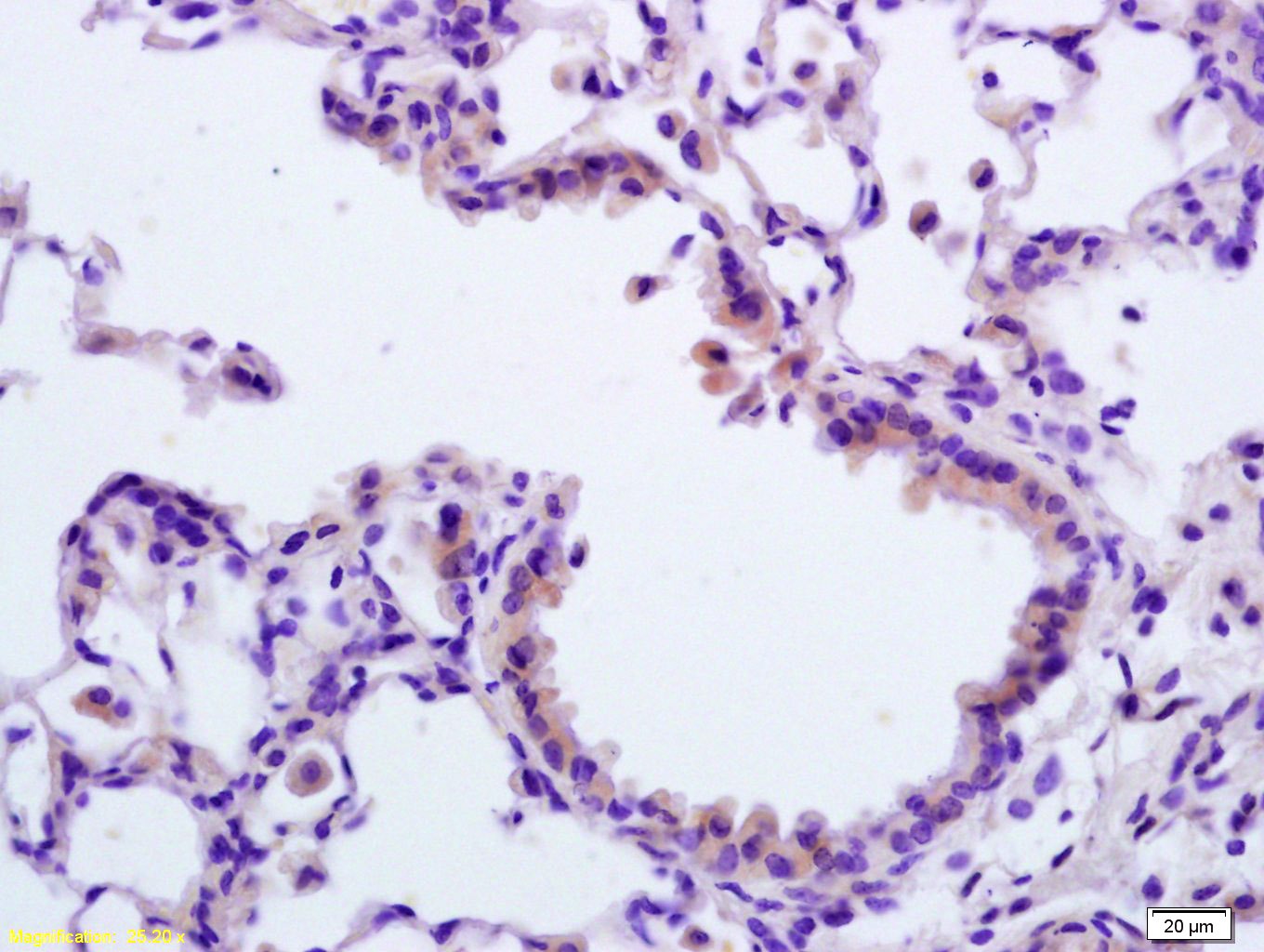 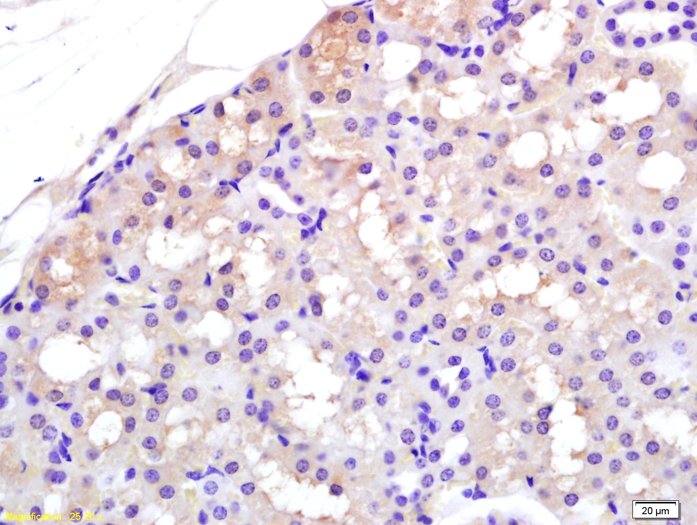 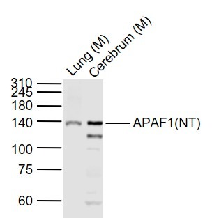 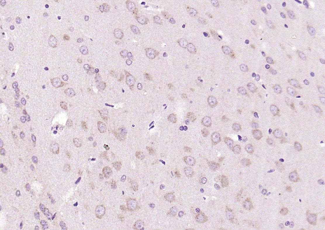 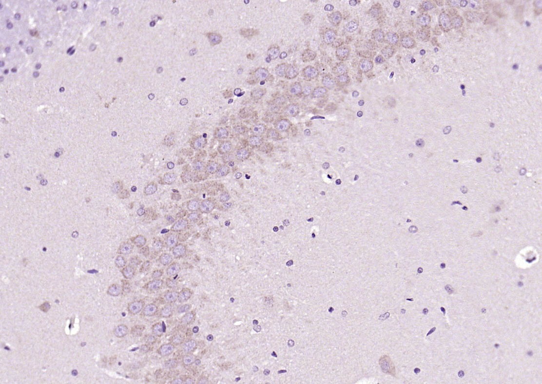 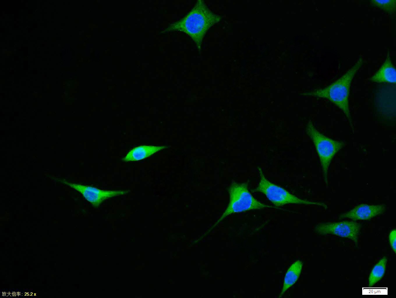 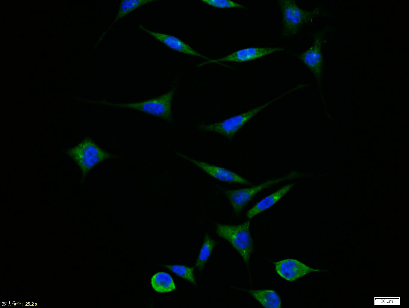 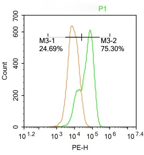 |
| 应用 | FCMICC/IFIFIHC-FrIHC-PWB |
| 推荐剂量 | FCM=0.2 μg/Test; ICC/IF=1:100-500; IF=1:100-500; IHC-Fr=1:100-500; IHC-P=1:100-500; WB=1:500-2000 |
| 抗体种类 | Polyclonal |
| 宿主来源 | Rabbit |
| 亚细胞定位 | Cytoplasm. |
| 组织特异性 | Ubiquitous. Highest levels of expression in adult spleen and peripheral blood leukocytes, and in fetal brain, kidney and lung. Isoform 1 is expressed in heart, kidney and liver. |
| 构建方式 | Polyclonal Antibody |
| 纯化方式 | Protein A purified |
| 性状 | Liquid |
| 缓冲液 | 0.01M TBS (pH7.4) with 1% BSA, 0.02% Proclin300 and 50% Glycerol. |
| 浓度 | 1 mg/mL |
| 研究背景 | This gene encodes a cytoplasmic protein that initiates apoptosis. This protein contains several copies of the WD-40 domain, a caspase recruitment domain (CARD), and an ATPase domain(NB-ARC). Upon binding cytochrome c and dATP, this protein forms an oligomeric apoptosome. The apoptosome binds and cleaves caspase 9 preproprotein, releasing its mature, activated form. Activated caspase 9 stimulates the subsequent caspase cascade that commits the cell to apoptosis. Alternative splicing results in several transcript variants encoding different isoforms. [provided by RefSeq, Jul 2008]. |
| 免疫原 | KLH conjugated synthetic peptide: human Apaf-1 |
| 抗原种属 | Human |
| 基因名称 | APAF1 |
| 基因ID | |
| 蛋白名称 | Apoptotic protease-activating factor 1 |
| Uniprot ID | |
| 研究领域 | Cytochrome C,Metabolism,Mitochondrial,Cytochrome C,Mitochondrial,Apoptosis,Mitochondrial markers |
| 功能 | Oligomeric Apaf-1 mediates the cytochrome c-dependent autocatalytic activation of pro-caspase-9 (Apaf-3), leading to the activation of caspase-3 and apoptosis. This activation requires ATP. Isoform 6 is less effective in inducing apoptosis. |
| 分子量 | Theoretical: 137 kDa. Actual: 140 kDa. |
| 储存方式 | Store at -20°C or -80°C for 12 months. Avoid repeated freeze-thaw cycles. |
| 运输方式 | Shipping with blue ice. |
 嗨!有任何问题?点我咨询
版权所有©2015-2026 TargetMol Chemicals Inc.保留所有权利.
沪ICP备20019793号-4 | 沪公网安备 31010602006700号 | 沪(静)应急管危经许[2024]203441