

购物车
 您的购物车当前为空


 还可以
还可以Anti-AKT1/2/3 Antibody (8U152) 是一种 Rabbit 抗体,靶向 AKT1/2/3。Anti-AKT1/2/3 Antibody (8U152) 可用于 FCM,ICC/IF,IHC,IP,WB。
还可以别名 STK-2, RAC-PK-gamma, RAC-PK-beta, RAC-PK-alpha, RAC-gamma serine/threonine-protein kinase, RAC-beta serine/threonine-protein kinase, RAC-alpha serine/threonine-protein kinase, Proto-oncogene c-Akt, Protein kinase B gamma, Protein kinase B beta, Protein kinase B alpha, Protein kinase B, Protein kinase Akt-3, Protein kinase Akt-2, PKBG, PKB RAC, PKB gamma, PKB beta, PKB alpha, PKB, EC 2.7.11.1, AKT3, AKT2, AKT1, AKT 3, AKT 2, AKT 1
Anti-AKT1/2/3 Antibody (8U152) 是一种 Rabbit 抗体,靶向 AKT1/2/3。Anti-AKT1/2/3 Antibody (8U152) 可用于 FCM,ICC/IF,IHC,IP,WB。
| 规格 | 价格 | 库存 | 数量 |
|---|---|---|---|
| 50 μL | ¥ 1,480 | 5日内发货 | |
| 100 μL | ¥ 2,485 | 5日内发货 |
TargetMol的所有产品仅用作科学研究或药证申报,不能被用于人体,我们不向个人提供产品和服务。请您遵守承诺用途,不得违反法律法规规定用于任何其他用途。
| 产品描述 | Anti-AKT1/2/3 Antibody (8U152) is a Rabbit antibody targeting AKT1/2/3. Anti-AKT1/2/3 Antibody (8U152) can be used in FCM,ICC/IF,IHC,IP,WB. |
| 别名 | STK-2, RAC-PK-gamma, RAC-PK-beta, RAC-PK-alpha, RAC-gamma serine/threonine-protein kinase, RAC-beta serine/threonine-protein kinase, RAC-alpha serine/threonine-protein kinase, Proto-oncogene c-Akt, Protein kinase B gamma, Protein kinase B beta, Protein kinase B alpha, Protein kinase B, Protein kinase Akt-3, Protein kinase Akt-2, PKBG, PKB RAC, PKB gamma, PKB beta, PKB alpha, PKB, EC 2.7.11.1, AKT3, AKT2, AKT1, AKT 3, AKT 2, AKT 1 |
| Ig Type | IgG |
| 克隆号 | 8U152 |
| 反应种属 | Human,Mouse,Rat |
| 验证活性 | 1. Western blot analysis of AKT1/2/3 on MCF-7 cell lysates using anti-AKT1/2/3 antibody at 1/1,000 dilution. 2. Immunohistochemical analysis of paraffin-embedded human kidney tissue using anti-AKT1/2/3 antibody. Counter stained with hematoxylin. 3. Immunohistochemical analysis of paraffin-embedded mouse brain tissue using anti-AKT1/2/3 antibody. Counter stained with hematoxylin. 4. Immunohistochemical analysis of paraffin-embedded mouse kidney tissue using anti-AKT1/2/3 antibody. Counter stained with hematoxylin. 5. Immunohistochemical analysis of paraffin-embedded mouse heart tissue using anti-AKT1/2/3 antibody. Counter stained with hematoxylin. 6. ICC staining AKT1/2/3 in A549 cells (green). The nuclear counter stain is DAPI (blue). Cells were fixed in paraformaldehyde, permeabilised with 0.25% Triton X100/PBS. 7. ICC staining AKT1/2/3 in CRC cells (green). The nuclear counter stain is DAPI (blue). Cells were fixed in paraformaldehyde, permeabilised with 0.25% Triton X100/PBS. 8. ICC staining AKT1/2/3 in SH-SY-5Y cells (green). The nuclear counter stain is DAPI (blue). Cells were fixed in paraformaldehyde, permeabilised with 0.25% Triton X100/PBS. 9. Flow cytometric analysis of A549 cells with AKT1/2/3 antibody at 1/50 dilution (blue) compared with an unlabelled control (cells without incubation with primary antibody; red). Alexa Fluor 488-conjugated goat anti rabbit IgG was used as the secondary antibody. 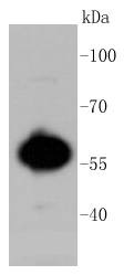 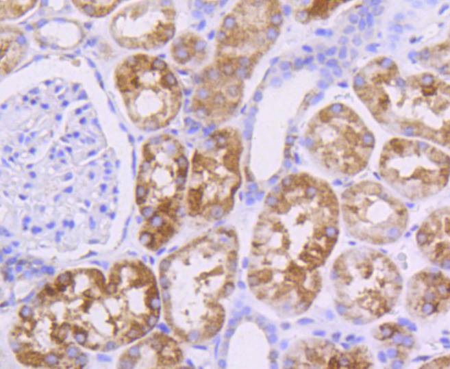 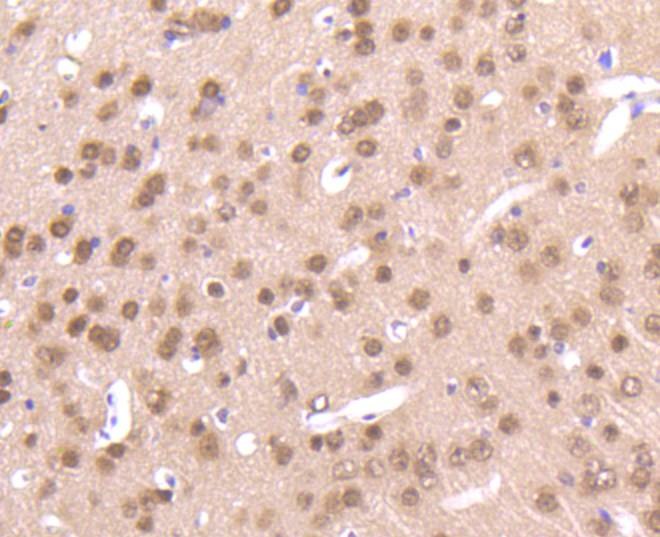 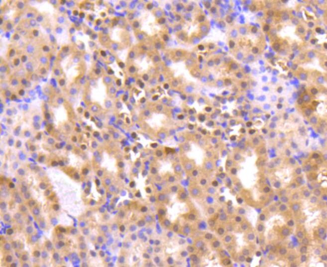 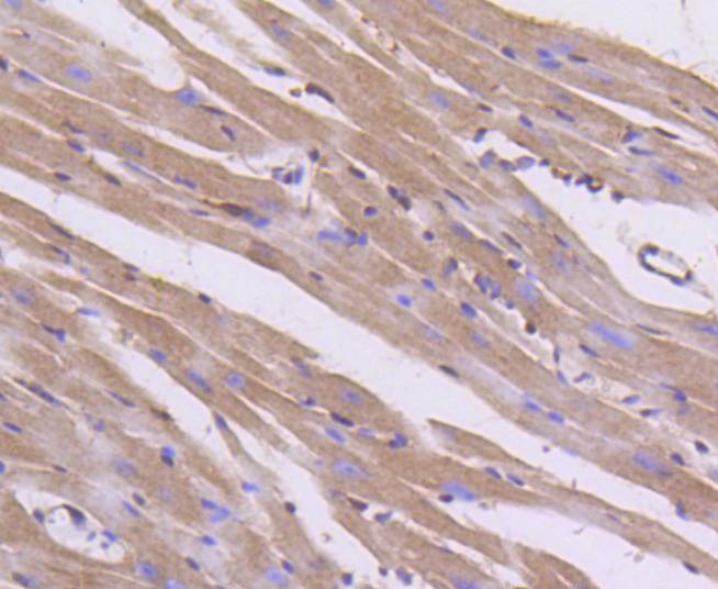 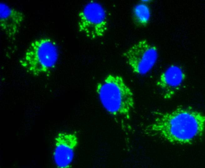 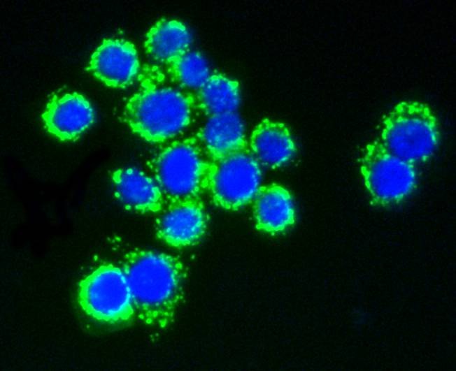 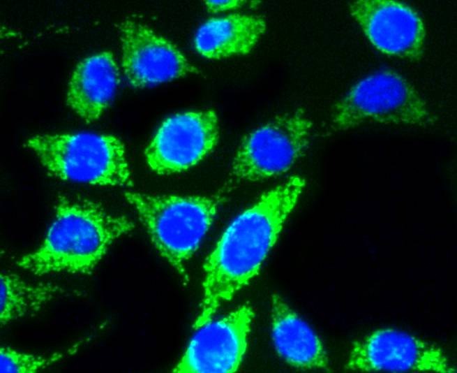 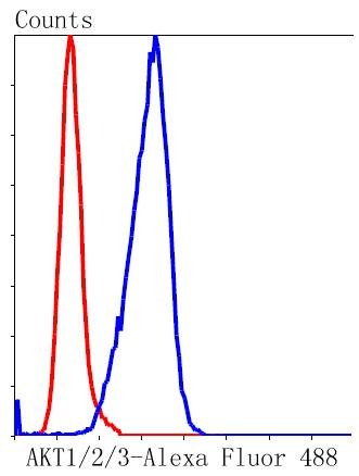 |
| 应用 | FCMICC/IFIHCIPWB |
| 推荐剂量 | WB: 1:1000-5000; IHC: 1:50-200; ICC/IF: 1:50-200; FCM: 1:50-100 |
| 抗体种类 | Monoclonal |
| 宿主来源 | Rabbit |
| 构建方式 | Recombinant Antibody |
| 纯化方式 | ProA affinity purified |
| 性状 | Liquid |
| 缓冲液 | 1*TBS (pH7.4), 1%BSA, 40%Glycerol. Preservative: 0.05% Sodium Azide. |
| 研究背景 | The serine/threonine kinase Akt family contains several members, including Akt1 (also designated PKB or RacPK), Akt2 (also designated PKBβ or RacPK-β) and Akt 3 (also designated PKBγ or thyoma viral proto-oncogene 3), which exhibit sequence homology with the protein kinase A and C families and are encoded by the c-Akt proto-oncogene. All members of the Akt family have a pleckstrin homology domain. Akt1 and Akt2 are activated by PDGF stimulation. This activation is dependent on PDGFR-β tyrosine residues 740 and 751, which bind the subunit of the phosphatidylinositol 3-kinase (PI 3-kinase) complex. Activation of Akt1 by insulin or insulin-growth factor-1(IGF-1) results in phosphorylation of both Thr 308 and Ser 473. Phosphorylation of both residues is important to generate a high level of Akt1 activity, and the phosphorylation of Thr 308 is not dependent on phosphorylation of Ser 473 in vivo. Thus, Akt proteins become phosphorylated and activated in insulin/IGF-1-stimulated cells by an upstream kinase(s). The activation of Akt1 and Akt2 is inhibited by the PI kinase inhibitor wortmannin, suggesting that the protein signals downstream of the PI kinases. |
| 偶联 | Unconjugated |
| 免疫原 | Recombinant Protein |
| Uniprot ID |
| 分子量 | Theoretical: 56 kDa. |
| 储存方式 | Store at -20°C or -80°C for 12 months. Avoid repeated freeze-thaw cycles. |
| 运输方式 | Shipping with blue ice. |
 嗨!有任何问题?点我咨询
版权所有©2015-2026 TargetMol Chemicals Inc.保留所有权利.
沪ICP备20019793号-4 | 沪公网安备 31010602006700号 | 沪(静)应急管危经许[2024]203441