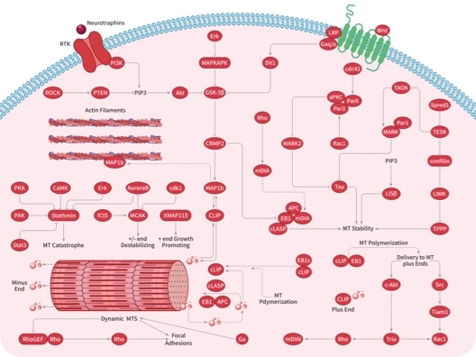
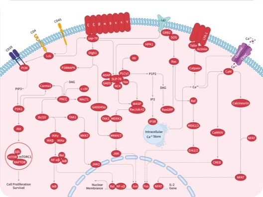
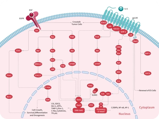
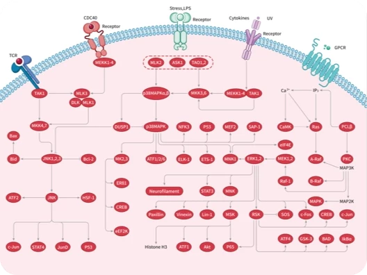
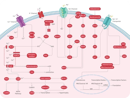
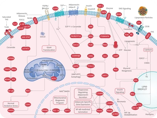
Mca-PLAC(p-OMeBz)-WAR(Dpa)-NH2 is a fluorogenic substrate for matrix metalloproteinase-14 (MMP-14). Upon cleavage by MMP-14, 7-methoxycoumarin-4-acetyl (Mca) is released and its fluorescence can be used to quantify MMP activity. Mca displays excitation emission maxima of 328 420 nm, respectively.
Mca-Ala-Pro-Lys(Dnp)-OH, a specific quenched fluorogenic substrate for ACE2, enables the detection of ACE2 activity in various tissues including urinary, heart, and lung.
D-Ala-Lys-AMCA, a known substrate of proton-coupled oligopeptide transporter 1 (PEPT1), emits blue fluorescence and may be transported into Caco-2 cells and liver cancer cells.
L-Leu-AMC is a fluorogenic substrate for leucine aminopeptidase.1Upon enzymatic cleavage by leucine aminopeptidase, AMC is released and its fluorescence can be used to quantify leucine aminopeptidase activity. AMC displays excitation emission maxima of 340-360 440-460 nm, respectively. 1.Izquierdo, M., Lin, D., O’Neill, S., et al.Development of a high-throughput screening assay to identify inhibitors of the major M17-leucyl aminopeptidase from Trypanosoma cruzi using rapidfire mass spectrometrySLAS Discov.25(9)1064-1071(2020)
Z-(L-Arg)-AMC is a fluorogenic substrate for trypsin, cathepsin B, and cathepsin H.1,2Upon enzymatic cleavage by trypsin, cathepsin B, or cathepsin H, 7-amino-4-methylcoumarin (AMC) is released and its fluorescence can be used to quantify trypsin, cathepsin B, and cathepsin H activity. AMC displays excitation emission maxima of 340-360 440-460 nm, respectively. 1.Zimmerman, M., Ashe, B., Yurewicz, E.C., et al.Sensitive assays for trypsin, elastase, and chymotrypsin using new fluorogenic substratesAnal. Biochem.78(1)47-51(1977) 2.Brindley, P.J., Kalinna, B.H., Dalton, J.P., et al.Proteolytic degradation of host hemoglobin by schistosomesMol. Biochem. Parasitol.89(1)1-9(1997)
Proteinase 3 (PR3, myeloblastin) is a polymorphonuclear leukocyte serine proteinase that degrades matrix proteins including fibronectin, laminin, vitronectin, and collagen type IV to generate antimicrobial peptides. Neutrophil elastase is a serine proteinase that is secreted by neutrophils during inflammation to destroy pathogens. Evaluating these enzymes is helpful to understanding inflammatory autoimmune processes. MeOSuc-AAPV-pNA is a highly sensitive peptide substrate that is hydrolyzed by both human and mouse neutrophil elastase and PR3, but not cathepsin G or chymotrypsin. Enzyme activity can be quantified by colorimetric detection of free p-nitroanilide at 405 nm.
Ac-ANW-AMC is a fluorogenic substrate for the β5i LMP7 subunit of the 20S immunoproteasome. [1] Upon cleavage, 7-amino-4-methylcoumarin (AMC) is released and its fluorescence can be used to quantify the activity of the β5i LMP7 subunit of the 20S immunoproteasome. AMC displays excitation emission maxima of 340-360 440-460 nm, respectively. Reference[1]. Winter, M.B., La Greca, F., Arastu-Kapur, S., et al. Immunoproteasome functions explained by divergence in cleavage specificity and regulation. eLife 6:e27364, (2017).
4-Methylumbelliferyl-β-D-galactoside (MUG) is a fluorogenic substrate for β-galactosidase (β-gal).1Hydrolysis of MUG by β-gal releases the fluorescent moiety 4-MU, which displays a pH-dependent excitation maximum of 330, 370, and 385 nm at pH 4.6, 7.4, and 10.4, respectively, and an emission maximum between 445-454 nm.2MUG has been used to detect β-gal activity in intact bacteria, yeast, and mammalian cells.1 1.Vidal-Aroca, F., Giannattasio, M., Brunelli, E., et al.One-step high-throughput assay for quantitative detection of β-galactosidase activity in intact Gram-negative bacteria, yeast, and mammalian cellsBiotechniques40(4)433-434(2006) 2.Zhi, H., Wang, J., Wang, S., et al.Fluorescent properties of hymecromone and fluorimetric analysis of hymecromone in compound dantong capsuleJ. Spectrosc.1(1)147128(2013)