
购物车
 您的购物车当前为空

 很棒
很棒纯度: 99.97%
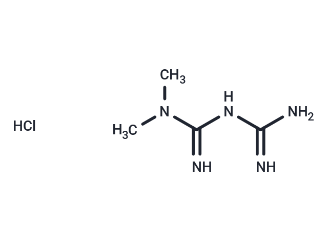
别名 盐酸二甲双胍, Metformin HCl, 1,1-Dimethylbiguanide hydrochloride, 1, 1-Dimethylbiguanide hydrochloride
Metformin hydrochloride (1,1-Dimethylbiguanide hydrochloride) 是一种 AMPK 激活剂,具有血脑屏障渗透性。Metformin hydrochloride 可通过提高胰岛素敏感性和减少肠道对葡萄糖的吸收来改善血糖控制,常用于 2 型糖尿病的研究。
 很棒
很棒纯度: 99.97%
Metformin hydrochloride (1,1-Dimethylbiguanide hydrochloride) 是一种 AMPK 激活剂,具有血脑屏障渗透性。Metformin hydrochloride 可通过提高胰岛素敏感性和减少肠道对葡萄糖的吸收来改善血糖控制,常用于 2 型糖尿病的研究。
| 规格 | 价格 | 库存 | 数量 |
|---|---|---|---|
| 50 mg | ¥ 168 | 现货 | |
| 100 mg | ¥ 233 | 现货 | |
| 500 mg | ¥ 536 | 现货 | |
| 1 g | ¥ 728 | 现货 | |
| 5 g | ¥ 2,160 | 现货 |
| 产品描述 | Metformin hydrochloride (1,1-Dimethylbiguanide hydrochloride) is an AMPK activator with blood-brain barrier permeability. Metformin hydrochloride may improve glycemic control by increasing insulin sensitivity and decreasing intestinal glucose uptake, and is commonly used in type 2 diabetes research. |
| 靶点活性 | MYCN-2 cells:> 100 mM, SK-N-Be2c cells:> 100 mM, HepG2 cells:98 mM, MDA-MB-231 cells:149 mM |
| 体外活性 | 方法: 卵巢癌细胞 A2780 和 SKOV3 用 Metformin hydrochloride (0.001-50 mM) 处理 24-48 h,使用 MTS 方法检测细胞活力。 |
| 体内活性 | 方法: 为建立 Metformin 诱导腹泻的模型,将 Metformin hydrochloride (125-500 mg/kg) 口服给药给健康和糖尿病肥胖 db/db 的 C57BL/6J 小鼠,每天两次,持续十三天。 |
| 细胞实验 | Hepatocytes were isolated from male Sprague Dawley (SD) rats by collagenase digestion. For the AMPK assay, cells were seeded in six-well plates at 1.5 × 10^6 cells/well in DMEM containing 100 U/ml penicillin, 100 μg/ml streptomycin, 10% FBS, 100 nM insulin, 100 nM dexamethasone, and 5 μg/ml transferrin for 4 hours. Cells were then cultured in serum-free DMEM for 16 hours followed by treatment for 1 hour or 7 hours with control medium, 5-aminoimidazole carboxamide riboside (AICAR), or metformin at concentrations indicated. For a 39-hour treatment, cells for both control and metformin (10 or 20 μM) groups were cultured in DMEM plus 5% FBS and 100 nM insulin, and the fresh control and metformin-containing medium were replaced every 12 hours (last medium change was 3 hours before harvest). After treatment, the cells were directly lysed in digitonin-containing and phosphatase inhibitor–containing buffer A, followed by precipitation with ammonium sulfate at 35% saturation. AMPK activity was determined by measurement of phosphorylation of a synthetic peptide substrate, SAMS (HMRSAMSGLHLVKRR). For ACC assay, the 35% ammonium sulfate precipitate from digitonin-lysed hepatocytes (4 μg each) was used for determination of ACC activity via 14CO2 fixation in the presence of 20 mM citrate as done previously. For fatty acid oxidation, the oxidation of 14C-oleate to acid-soluble products was performed as done previously, but in medium M199 in the absence of albumin [1]. |
| 动物实验 | Oral gavage was used to administer 1 ml of metformin (100 mg/ml) or water alone to male SD rats (300–350 g, n = 7–8). Rats were treated once or twice a day for 5 days. Rats were starved for 20 hours and then re-fed for 2 hours before the final dose; 4 hours after the final dose, the animals were anesthetized and livers rapidly removed by freeze clamping followed by blood withdrawal. RNA was prepared from the freeze-clamped liver by RNA isolation reagent. Nuclear extracts were prepared from a pool of seven rat livers. Glucose levels were determined using the standard glucose oxidase assay kit; β-hydroxybutyrate concentrations were assayed by measuring the reduction of NAD to NADH with a standard assay kit. FFA levels were measured with the assay kit [1]. MCF10A-ER-Src cells (5 × 10^6) were injected into the right flank of 18 female nu/nu mice, all of which developed tumors in 10 d with a size of ~100 mm^3. The mice were randomly distributed into six groups (three mice/group) that were untreated or treated by intratumoral injections every 5 d (four cycles) with 1 mg/kg or 4 mg/kg doxorubicin, 200 μg/mL metformin (diluted in the drinking water), or the combination. In another experiment, LNCaP and DU145 prostate cancer cells (5 × 10^6) were injected into the right flank of 12 female nu/nu mice, all of which developed tumors in 10 d with a size of ~75 mm^3. The mice were randomly distributed into four groups that were untreated or treated by intratumoral injections every 5 d (four cycles) with 4 mg/kg doxorubicin and/or 200 μg/mL metformin. In another experiment, A375 and MDA-MB-435 melanoma cells (7 × 10^6) were injected into the right flank of 12 female nu/nu mice, all of which developed tumors in 10 d with a size of ~50 mm3. The mice were randomly distributed into four groups that were untreated or treated by intratumoral injections every 5 d (four cycles) with 10 mg/kg cisplatin and/or 200 μg/mL metformin.Finally, SNU-449 liver cancer cells (10^7) were injected into the right flank of 12 female nu/nu mice, all of which developed tumors in 10 d with a size of ~50 mm^3. The mice were randomly distributed into four groups that were untreated or treated by intratumoral injections every 5 d (four cycles) with 10 mg/kg cisplatin and/or 200 μg/mL metformin. Tumor volume (mean ± SD) was measured at various times after the initial injection [3]. |
| 别名 | 盐酸二甲双胍, Metformin HCl, 1,1-Dimethylbiguanide hydrochloride, 1, 1-Dimethylbiguanide hydrochloride |
| 分子量 | 165.63 |
| 分子式 | C4H12ClN5 |
| CAS No. | 1115-70-4 |
| Smiles | Cl.CN(C)C(=N)NC(N)=N |
| 密度 | 1.36. Temperature:20.1 °C. |
| 颜色 | White |
| 物理性状 | Solid |
| 存储 | Powder: -20°C for 3 years | In solvent: -80°C for 1 year | Shipping with blue ice/Shipping at ambient temperature. | |||||||||||||||||||||||||||||||||||
| 溶解度信息 | H2O: 193.21 mM, Sonication is recommended. DMSO: 50 mg/mL (301.88 mM), Sonication is recommended. | |||||||||||||||||||||||||||||||||||
溶液配制表 | ||||||||||||||||||||||||||||||||||||
H2O/DMSO
| ||||||||||||||||||||||||||||||||||||
 比如您的给药剂量是10 mg/kg,每只动物体重20g,给药体积100 μL, 一共给药动物10只,您使用的配方为5%
 DMSO + 30%PEG300 + 5%Tween 80 + 60%Saline/PBS/ddH2O, 那么您的工作液浓度为2mg/mL。
 以上为“体内实验配液计算器”的使用方法举例,并不是具体某个化合物的推荐配制方式,请根据您的实验动物和给药方式选择适当的溶解方案。
对于不同动物的给药剂量换算,您也可以参考 更多
 嗨!有任何问题?点我咨询
版权所有©2015-2026 TargetMol Chemicals Inc.保留所有权利.
沪ICP备20019793号-4 | 沪公网安备 31010602006700号 | 沪(静)应急管危经许[2024]203441