

购物车
 您的购物车当前为空


 还可以
还可以Anti-PIK3R1 Antibody (9O121) 是一种 Rabbit 抗体,靶向 PIK3R1。Anti-PIK3R1 Antibody (9O121) 可用于 FCM,ICC/IF,IHC,WB。
还可以别名 SH3_PI3K_p85alpha, PtdIns-3-kinase regulatory subunit p85-alpha, PtdIns-3-kinase regulatory subunit alpha, PI3-kinase subunit p85-alpha, PI3-kinase regulatory subunit alpha, PI3-kinase p85 subunit gamma, PI3-kinase p85 subunit alpha, PI3K regulatory subunit alpha, PI 3-kinase p85-α, PI 3-kinase p85α, PI 3-kinase p85 α, PI 3 Kinase p85 alpha, phosphoinositide-3-kinase regulatory subunit 1, Phosphatidylinositol 3-kinase regulatory subunit alpha, Phosphatidylinositol 3-kinase 85 kDa regulatory subunit alpha, p85-ALPHA, P85A, p85, IMD36, GRB1, AGM7
Anti-PIK3R1 Antibody (9O121) 是一种 Rabbit 抗体,靶向 PIK3R1。Anti-PIK3R1 Antibody (9O121) 可用于 FCM,ICC/IF,IHC,WB。
| 规格 | 价格 | 库存 | 数量 |
|---|---|---|---|
| 50 μL | ¥ 1,495 | 5日内发货 | |
| 100 μL | ¥ 2,480 | 5日内发货 |
TargetMol的所有产品仅用作科学研究或药证申报,不能被用于人体,我们不向个人提供产品和服务。请您遵守承诺用途,不得违反法律法规规定用于任何其他用途。
| 产品描述 | Anti-PIK3R1 Antibody (9O121) is a Rabbit antibody targeting PIK3R1. Anti-PIK3R1 Antibody (9O121) can be used in FCM,ICC/IF,IHC,WB. |
| 别名 | SH3_PI3K_p85alpha, PtdIns-3-kinase regulatory subunit p85-alpha, PtdIns-3-kinase regulatory subunit alpha, PI3-kinase subunit p85-alpha, PI3-kinase regulatory subunit alpha, PI3-kinase p85 subunit gamma, PI3-kinase p85 subunit alpha, PI3K regulatory subunit alpha, PI 3-kinase p85-α, PI 3-kinase p85α, PI 3-kinase p85 α, PI 3 Kinase p85 alpha, phosphoinositide-3-kinase regulatory subunit 1, Phosphatidylinositol 3-kinase regulatory subunit alpha, Phosphatidylinositol 3-kinase 85 kDa regulatory subunit alpha, p85-ALPHA, P85A, p85, IMD36, GRB1, AGM7 |
| Ig Type | IgG |
| 克隆号 | 9O121 |
| 反应种属 | Human,Mouse,Rat |
| 验证活性 | 1. Western blot analysis of PI 3 Kinase p85 alpha on different lysates using anti-PI 3 Kinase p85 alpha antibody at 1/1,000 dilution. Positive control: Lane 1: MCF-7, Lane 2: Raji. 2. ICC staining PI 3 Kinase p85 alpha in Hela cells (red). The nuclear counter stain is DAPI (blue). Cells were fixed in paraformaldehyde, permeabilised with 0.25% Triton X100/PBS. 3. ICC staining PI 3 Kinase p85 alpha in MCF-7 cells (red). The nuclear counter stain is DAPI (blue). Cells were fixed in paraformaldehyde, permeabilised with 0.25% Triton X100/PBS. 4. ICC staining PI 3 Kinase p85 alpha in HepG2 cells (red). The nuclear counter stain is DAPI (blue). Cells were fixed in paraformaldehyde, permeabilised with 0.25% Triton X100/PBS. 5. ICC staining PI 3 Kinase p85 alpha in NIH/3T3 cells (red). The nuclear counter stain is DAPI (blue). Cells were fixed in paraformaldehyde, permeabilised with 0.25% Triton X100/PBS. 6. Flow cytometric analysis of HepG2 cells with PI 3 Kinase p85 alpha antibody at 1/50 dilution (red) compared with an unlabelled control (cells without incubation with primary antibody; black). Alexa Fluor 488-conjugated goat anti rabbit IgG was used as the secondary antibody. 7. Immunohistochemical analysis of paraffin-embedded human liver carcinoma tissue using anti-PI 3 Kinase p85 alpha antibody. The section was pre-treated using heat mediated antigen retrieval with Tris-EDTA buffer (pH 8.0-8.4) for 20 minutes.The tissues were blocked in 5% BSA for 30 minutes at room temperature, washed with ddH2O and PBS, and then probed with the primary antibody (1/50) for 30 minutes at room temperature. The detection was performed using an HRP conjugated compact polymer system. DAB was used as the chromogen. Tissues were counterstained with hematoxylin and mounted with DPX. 8. Immunohistochemical analysis of paraffin-embedded human kidney tissue using anti-PI 3 Kinase p85 alpha antibody. The section was pre-treated using heat mediated antigen retrieval with Tris-EDTA buffer (pH 8.0-8.4) for 20 minutes.The tissues were blocked in 5% BSA for 30 minutes at room temperature, washed with ddH2O and PBS, and then probed with the primary antibody (1/50) for 30 minutes at room temperature. The detection was performed using an HRP conjugated compact polymer system. DAB was used as the chromogen. Tissues were counterstained with hematoxylin and mounted with DPX 9. Immunohistochemical analysis of paraffin-embedded mouse kidney tissue using anti-PI 3 Kinase p85 alpha antibody. The section was pre-treated using heat mediated antigen retrieval with Tris-EDTA buffer (pH 8.0-8.4) for 20 minutes.The tissues were blocked in 5% BSA for 30 minutes at room temperature, washed with ddH2O and PBS, and then probed with the primary antibody (1/50) for 30 minutes at room temperature. The detection was performed using an HRP conjugated compact polymer system. DAB was used as the chromogen. Tissues were counterstained with hematoxylin and mounted with DPX. 10. Immunohistochemical analysis of paraffin-embedded mouse heart tissue using anti-PI 3 Kinase p85 alpha antibody. The section was pre-treated using heat mediated antigen retrieval with Tris-EDTA buffer (pH 8.0-8.4) for 20 minutes.The tissues were blocked in 5% BSA for 30 minutes at room temperature, washed with ddH2O and PBS, and then probed with the primary antibody (1/50) for 30 minutes at room temperature. The detection was performed using an HRP conjugated compact polymer system. DAB was used as the chromogen. Tissues were counterstained with hematoxylin and mounted with DPX. 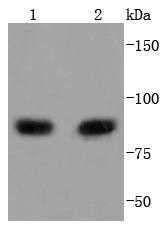 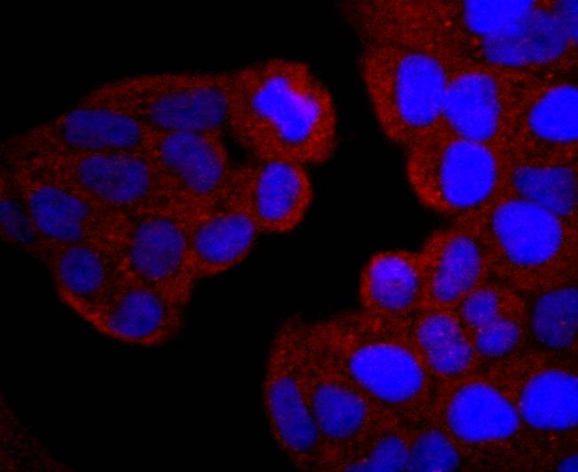 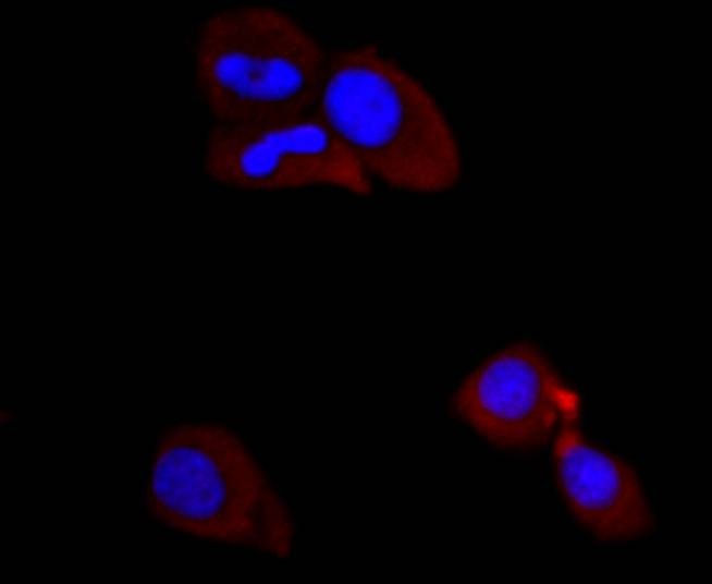 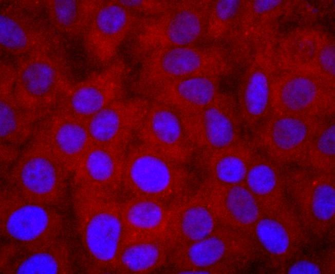 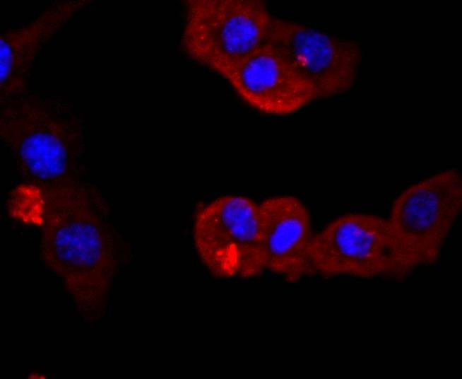 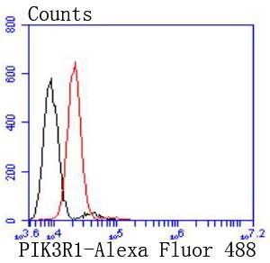 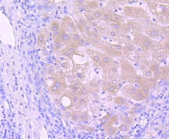 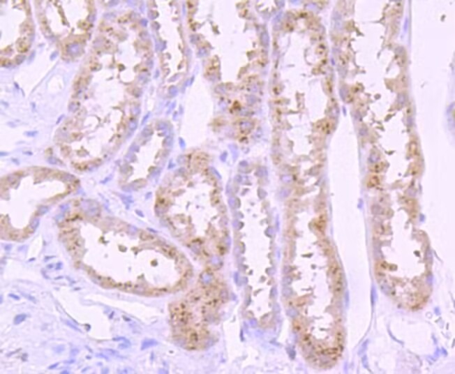 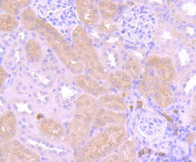 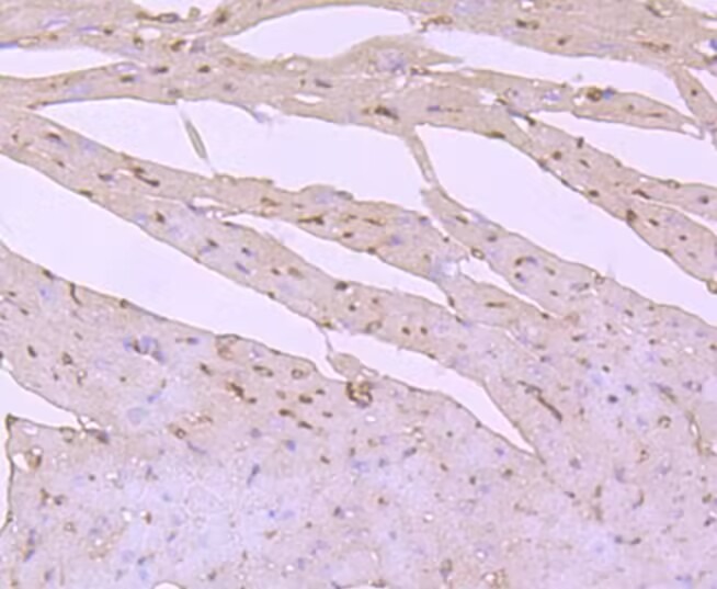 |
| 应用 | FCMICC/IFIHCWB |
| 推荐剂量 | WB: 1:1000-2000; IHC: 1:50-200; ICC/IF: 1:50-200; FCM: 1:50-100 |
| 抗体种类 | Monoclonal |
| 宿主来源 | Rabbit |
| 构建方式 | Recombinant Antibody |
| 纯化方式 | ProA affinity purified |
| 性状 | Liquid |
| 缓冲液 | 1*TBS (pH7.4), 0.05% BSA, 40% Glycerol. Preservative: 0.05% Sodium Azide. |
| 研究背景 | Phosphatidylinositol 3-kinase (PI 3-kinase) phosphorylates the 3' OH position of the inositol ring of inositol lipids and is composed of p85 and p110 subunits. PI 3-kinase p85 lacks PI 3-kinase activity and acts as an adapter, coupling p110 to activated protein tyrosine kinase. Two forms of p85 have been described (p85α and p85β), each possessing one SH3 and two SH2 domains. PI 3-kinase p85α, also known as GRB1, phosphatidylinositol 3-kinase regulatory 1 or p85, is a 724 amino acid protein that exists as four alternatively spliced isoforms. Involved in insulin metabolism, defects in the PI 3-kinase p85α gene have been linked to insulin resistance. PI 3-kinase p85α is polyubiquitinated in T-cells by Cbl-b, and has multiple phosphorylated amino acid residues, including a phosphorylated tyrosine residue at position 467. |
| 偶联 | Unconjugated |
| 免疫原 | A synthesized peptide: C-terminal human PI 3 Kinase p85 alpha |
| 抗原种属 | Human |
| Uniprot ID |
| 分子量 | Theoretical: 84 kDa. |
| 储存方式 | Store at -20°C or -80°C for 12 months. Avoid repeated freeze-thaw cycles. |
| 运输方式 | Shipping with blue ice. |
 嗨!有任何问题?点我咨询
版权所有©2015-2026 TargetMol Chemicals Inc.保留所有权利.
沪ICP备20019793号-4 | 沪公网安备 31010602006700号 | 沪(静)应急管危经许[2024]203441