

购物车
 您的购物车当前为空


 还可以
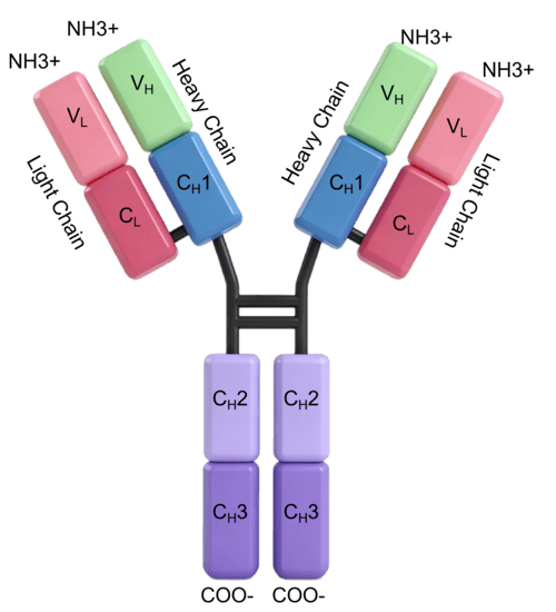
还可以Anti-CD63 Antibody (4G617) 是一种 Mouse 抗体,靶向 CD63。Anti-CD63 Antibody (4G617) 可用于 ELISA,FCM,IF,IHC,WB。
还可以别名 Tspan30, ME491, CD63 molecule, C75951
Anti-CD63 Antibody (4G617) 是一种 Mouse 抗体,靶向 CD63。Anti-CD63 Antibody (4G617) 可用于 ELISA,FCM,IF,IHC,WB。
| 规格 | 价格 | 库存 | 数量 |
|---|---|---|---|
| 50 μL | ¥ 1,300 | 5日内发货 | |
| 100 μL | ¥ 2,195 | 5日内发货 |
TargetMol的所有产品仅用作科学研究或药证申报,不能被用于人体,我们不向个人提供产品和服务。请您遵守承诺用途,不得违反法律法规规定用于任何其他用途。
| 产品描述 | Anti-CD63 Antibody (4G617) is a Mouse antibody targeting CD63. Anti-CD63 Antibody (4G617) can be used in ELISA,FCM,IF,IHC,WB. |
| 别名 | Tspan30, ME491, CD63 molecule, C75951 |
| Ig Type | IgG1 |
| 克隆号 | 4G617 |
| 反应种属 | Human, Rabbit |
| 验证活性 | 1. Western Blot -Positive WB detected in: A549 whole cell lysate, Hela whole cell lysate, HepG2 whole cell lysate, MCF-7 whole cell lysate -All lanes CD63 antibody at 1:1000 -Secondary: Goat polyclonal to mouse IgG at 1/50000 dilution -Predicted band size: 30-120 KD KDa -Observed band size: 30-120 KD KDa -Exposure time:1min 2. Western Blot -Positive WB detected in: Raji whole cell lysate -All lanes CD63 antibody at 1:1000 -Secondary: Goat polyclonal to mouse IgG at 1/50000 dilution -Predicted band size: 30-120 KD KDa -Observed band size: 30-120 KD KDa -Exposure time:1min 3. Western Blot -Positive WB detected in: A375 whole cell lysate, Rabbit spleen tissue -All lanes CD63 antibody at 1:1000 -Secondary: Goat polyclonal to mouse IgG at 1/50000 dilution -Predicted band size: 30-120 KD KDa -Observed band size: 30-120 KD KDa -Exposure time:1min` 4. IHC image of TMAH-00215 diluted at 1:500 and staining in paraffin-embedded human lung cancer tissue performed on a Leica BondTM system. After dewaxing and hydration, antigen retrieval was mediated by high pressure in a citrate buffer (pH 6.0). Section was blocked with 10% normal goat serum 30min at 37°C. Then primary antibody (1% BSA) was incubated at 4°C overnight. The primary is detected by a Goat anti-rabbit IgG labeled by HRP and visualized using 0.05% DAB. 5. IHC image of TMAH-00215 diluted at 1:500 and staining in paraffin-embedded human lung cancer tissue performed on a Leica BondTM system. After dewaxing and hydration, antigen retrieval was mediated by high pressure in a citrate buffer (pH 6.0). Section was blocked with 10% normal goat serum 30min at 37°C. Then primary antibody (1% BSA) was incubated at 4°C overnight. The primary is detected by a Goat anti-rabbit IgG labeled by HRP and visualized using 0.05% DAB. 6. IHC image of TMAH-00215 diluted at 1:500 and staining in paraffin-embedded human lung cancer tissue performed on a Leica BondTM system. After dewaxing and hydration, antigen retrieval was mediated by high pressure in a citrate buffer (pH 6.0). Section was blocked with 10% normal goat serum 30min at 37°C. Then primary antibody (1% BSA) was incubated at 4°C overnight. The primary is detected by a Goat anti-rabbit IgG labeled by HRP and visualized using 0.05% DAB. 7. Immunofluorescence staining of A549 cells with TMAH-00215 at 1:50, counter-stained with DAPI. The cells were fixed in 4% formaldehyde and blocked in 10% normal Goat Serum. The cells were then incubated with the antibody overnight at 4°C. Nuclear DNA was labeled in blue with DAPI. The secondary antibody was FITC-conjugated AffiniPure Goat Anti-Mouse IgG (H+L). 8. Immunofluorescence staining of Hela cells with TMAH-00215 at 1:50, counter-stained with DAPI. The cells were fixed in 4% formaldehyde and blocked in 10% normal Goat Serum. The cells were then incubated with the antibody overnight at 4°C. Nuclear DNA was labeled in blue with DAPI. The secondary antibody was FITC-conjugated AffiniPure Goat Anti-Mouse IgG (H+L). 9. Immunofluorescence staining of MCF-7 cells with TMAH-00215 at 1:50, counter-stained with DAPI. The cells were fixed in 4% formaldehyde and blocked in 10% normal Goat Serum. The cells were then incubated with the antibody overnight at 4°C. Nuclear DNA was labeled in blue with DAPI. The secondary antibody was FITC-conjugated AffiniPure Goat Anti-Mouse IgG (H+L). 10. Overlay histogram showing A549 cells stained with TMAH-00215 (red line) at 1:100. The cells were fixed in 4% formaldehyde and permeated by 0.2% TritonX-100. Then 10% normal goat serum was Incubated to block non-specific protein-protein interactions followed by the antibody (1µg/1*10^6 cells) for 1 h at 4°C. The secondary antibody used was FITC-conjugated Goat Anti-Mouse IgG(H+L) at 1/100 dilution for 30min at 4°C. Isotype control antibody (green line) was mouse IgG2b (1µg/1*106cells) used under the same conditions. Acquisition of >10,000 events was performed. 11. Overlay histogram showing Hela cells stained with TMAH-00215 (red line) at 1:100. The cells were fixed in 4% formaldehyde and permeated by 0.2% TritonX-100. Then 10% normal goat serum was Incubated to block non-specific protein-protein interactions followed by the antibody (1µg/1*10^6 cells) for 1 h at 4°C. The secondary antibody used was FITC-conjugated Goat Anti-Mouse IgG(H+L) at 1/100 dilution for 30min at 4°C. Isotype control antibody (green line) was mouse IgG2b (1µg/1*106cells) used under the same conditions. Acquisition of >10,000 events was performed. 12. Overlay histogram showing HepG2 cells stained with TMAH-00215 (red line) at 1:100. The cells were fixed in 4% formaldehyde and permeated by 0.2% TritonX-100. Then 10% normal goat serum was Incubated to block non-specific protein-protein interactions followed by the antibody (1µg/1*10^6 cells) for 1 h at 4°C. The secondary antibody used was FITC-conjugated Goat Anti-Mouse IgG(H+L) at 1/100 dilution for 30min at 4°C. Isotype control antibody (green line) was mouse IgG2b (1µg/1*106cells) used under the same conditions. Acquisition of >10,000 events was performed. 13. Overlay histogram showing K562 cells stained with TMAH-00215 (red line) at 1:100. The cells were fixed in 4% formaldehyde and permeated by 0.2% TritonX-100. Then 10% normal goat serum was Incubated to block non-specific protein-protein interactions followed by the antibody (1µg/1*10^6 cells) for 1 h at 4°C. The secondary antibody used was FITC-conjugated Goat Anti-Mouse IgG(H+L) at 1/100 dilution for 30min at 4°C. Isotype control antibody (green line) was mouse IgG2b (1µg/1*106cells) used under the same conditions. Acquisition of >10,000 events was performed. 14. 1-Exosomes extracted from Hela cells 2-Exosomes extracted from Hela cells 3-Exosomes extracted from urine |
| 应用 | ELISAFCMIFIHCWB |
| 抗体种类 | Monoclonal |
| 宿主来源 | Mouse |
| 亚细胞定位 | Cell membrane; Multi-pass membrane protein. Lysosome membrane; Multi-pass membrane protein. Late endosome membrane; Multi-pass membrane protein. Endosome, multivesicular body. Melanosome. Secreted, extracellular exosome. Cell surface. |
| 组织特异性 | Detected in platelets (at protein level). Dysplastic nevi, radial growth phase primary melanomas, hematopoietic cells, tissue macrophages. |
| 构建方式 | Hybridoma Monoclonal Antibody |
| 纯化方式 | Protein G purified |
| 性状 | Liquid |
| 缓冲液 | Preservative: 0.03% Proclin 300. Constituents: 50% Glycerol, 0.01M PBS, PH 7.4. |
| 纯度 | >95% |
| 研究背景 | Functions as cell surface receptor for TIMP1 and plays a role in the activation of cellular signaling cascades. Plays a role in the activation of ITGB1 and integrin signaling, leading to the activation of AKT, FAK/PTK2 and MAP kinases. Promotes cell survival, reorganization of the actin cytoskeleton, cell adhesion, spreading and migration, via its role in the activation of AKT and FAK/PTK2. Plays a role in VEGFA signaling via its role in regulating the internalization of KDR/VEGFR2. Plays a role in intracellular vesicular transport processes, and is required for normal trafficking of the PMEL luminal domain that is essential for the development and maturation of melanocytes. Plays a role in the adhesion of leukocytes onto endothelial cells via its role in the regulation of SELP trafficking. May play a role in mast cell degranulation in response to Ms4a2/FceRI stimulation, but not in mast cell degranulation in response to other stimuli. |
| 偶联 | Unconjugated |
| 免疫原 | Recombinant Protein: Human CD63 antigen Protein (103-203AA) |
| 抗原种属 | Human |
| 基因名称 | CD63 |
| 基因ID | |
| Uniprot ID | |
| 研究领域 | Epigenetics and Nuclear Signaling |
| 储存方式 | Store at -20°C or -80°C for 12 months. Avoid repeated freeze-thaw cycles. |
| 运输方式 | Shipping with blue ice. |
 嗨!有任何问题?点我咨询
版权所有©2015-2026 TargetMol Chemicals Inc.保留所有权利.
沪ICP备20019793号-4 | 沪公网安备 31010602006700号 | 沪(静)应急管危经许[2024]203441