

购物车
 您的购物车当前为空


 还可以
还可以Anti-HADHSC Antibody (1K241) 是一种 Mouse 抗体,靶向 HADHSC。Anti-HADHSC Antibody (1K241) 可用于 ICC,IHC,WB。
还可以别名 Short-chain 3-hydroxyacyl-CoA dehydrogenase, short chain 3-hydroxyacyl-coa dehydrogenase, Short chain 3 hydroxyacyl CoA dehydrogenase mitochondrial, SCHAD, formerly, SCHAD, OTTHUMP00000219688, OTTHUMP00000162626, MSCHAD, mitochondrial, MGC8392, Medium and short-chain L-3-hydroxyacyl-coenzyme A dehydrogenase, Medium and short chain L 3 hydroxyacyl coenzyme A dehydrogenase, M SCHAD, L 3 hydroxyacyl Coenzyme A dehydrogenase short chain, hydroxyacyl-coenzyme A dehydrogenase, mitochondrial, Hydroxyacyl-coenzyme A dehydrogenase, Hydroxyacyl CoA dehydrogenase, HHF4, HCDH_HUMAN, HCDH, HADSC, formerly, HADHSC, formerly, HADHSC, HADH1, HADH, HAD, 3 hydroxyacyl Coenzyme A dehydrogenase
Anti-HADHSC Antibody (1K241) 是一种 Mouse 抗体,靶向 HADHSC。Anti-HADHSC Antibody (1K241) 可用于 ICC,IHC,WB。
| 规格 | 价格 | 库存 | 数量 |
|---|---|---|---|
| 50 μL | ¥ 1,985 | 5日内发货 | |
| 100 μL | ¥ 2,990 | 5日内发货 |
TargetMol的所有产品仅用作科学研究或药证申报,不能被用于人体,我们不向个人提供产品和服务。请您遵守承诺用途,不得违反法律法规规定用于任何其他用途。
| 产品描述 | Anti-HADHSC Antibody (1K241) is a Mouse antibody targeting HADHSC. Anti-HADHSC Antibody (1K241) can be used in ICC,IHC,WB. |
| 别名 | Short-chain 3-hydroxyacyl-CoA dehydrogenase, short chain 3-hydroxyacyl-coa dehydrogenase, Short chain 3 hydroxyacyl CoA dehydrogenase mitochondrial, SCHAD, formerly, SCHAD, OTTHUMP00000219688, OTTHUMP00000162626, MSCHAD, mitochondrial, MGC8392, Medium and short-chain L-3-hydroxyacyl-coenzyme A dehydrogenase, Medium and short chain L 3 hydroxyacyl coenzyme A dehydrogenase, M SCHAD, L 3 hydroxyacyl Coenzyme A dehydrogenase short chain, hydroxyacyl-coenzyme A dehydrogenase, mitochondrial, Hydroxyacyl-coenzyme A dehydrogenase, Hydroxyacyl CoA dehydrogenase, HHF4, HCDH_HUMAN, HCDH, HADSC, formerly, HADHSC, formerly, HADHSC, HADH1, HADH, HAD, 3 hydroxyacyl Coenzyme A dehydrogenase |
| 克隆号 | 1K241 |
| 反应种属 | Human,Mouse,zebrafish |
| 验证活性 | 1. Western blot analysis of HADH on different lysates with HADH at 1/1,000 dilution. Lane 1: HepG2 cell lysate (20 ug/Lane), Lane 2: HeLa cell lysate (20 ug/Lane), Lane 3: HT-29 cell lysate (20 ug/Lane), Lane 4: HL-60 cell lysate (20 ug/Lane), Lane 5: A431 cell lysate (20 ug/Lane), Lane 6: K-562 cell lysate (20 ug/Lane), Lane 7: Human liver tissue lysate (40 ug/Lane), Lane 8: Mouse liver tissue lysate (40 ug/Lane), Lane 9: Mouse heart tissue lysate (40 ug/Lane), Lane 10: Rat liver tissue lysate (40 ug/Lane), Lane 11: Rat heart tissue lysate (40 ug/Lane), Lane 12: Zebrafish tissue lysate (40 ug/Lane), Predicted band size: 34 kDa, Observed band size: 34 kDa. 2. Immunocytochemistry analysis of HeLa cells labeling HADH at 1/100 dilution. Cells were fixed in 4% paraformaldehyde for 20 minutes at room temperature, permeabilized with 0.1% Triton X-100 in PBS for 5 minutes at room temperature, then blocked with 1% BSA in 10% negative goat serum for 1 hour at room temperature. Cells were then incubated with HADH at 1/100 dilution in 1% BSA in PBST overnight at 4 ℃. Goat Anti-Mouse IgG H&L (iFluor 488) was used as the secondary antibody at 1/1,000 dilution. PBS instead of the primary antibody was used as the secondary antibody only control. Nuclear DNA was labelled in blue with DAPI. 3. Immunohistochemical analysis of paraffin-embedded human kidney tissue with HADH at 1/1,000 dilution.The section was pre-treated using heat mediated antigen retrieval with Tris-EDTA buffer (pH 9.0) for 20 minutes. The tissues were blocked in 1% BSA for 20 minutes at room temperature, washed with ddH2O and PBS, and then probed with the primary antibody at 1/1,000 dilution for 1 hour at room temperature. The detection was performed using an HRP conjugated compact polymer system. DAB was used as the chromogen. Tissues were counterstained with hematoxylin and mounted with DPX. 4. Immunohistochemical analysis of paraffin-embedded mouse liver tissue with HADH at 1/1,000 dilution. The section was pre-treated using heat mediated antigen retrieval with Tris-EDTA buffer (pH 9.0) for 20 minutes. The tissues were blocked in 1% BSA for 20 minutes at room temperature, washed with ddH2O and PBS, and then probed with the primary antibody at 1/1,000 dilution for 1 hour at room temperature. The detection was performed using an HRP conjugated compact polymer system. DAB was used as the chromogen. Tissues were counterstained with hematoxylin and mounted with DPX. 5. Immunohistochemical analysis of paraffin-embedded rat liver tissue with HADH at 1/1,000 dilution. The section was pre-treated using heat mediated antigen retrieval with Tris-EDTA buffer (pH 9.0) for 20 minutes. The tissues were blocked in 1% BSA for 20 minutes at room temperature, washed with ddH2O and PBS, and then probed with the primary antibody at 1/1,000 dilution for 1 hour at room temperature. The detection was performed using an HRP conjugated compact polymer system. DAB was used as the chromogen. Tissues were counterstained with hematoxylin and mounted with DPX. 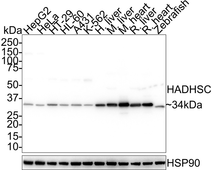 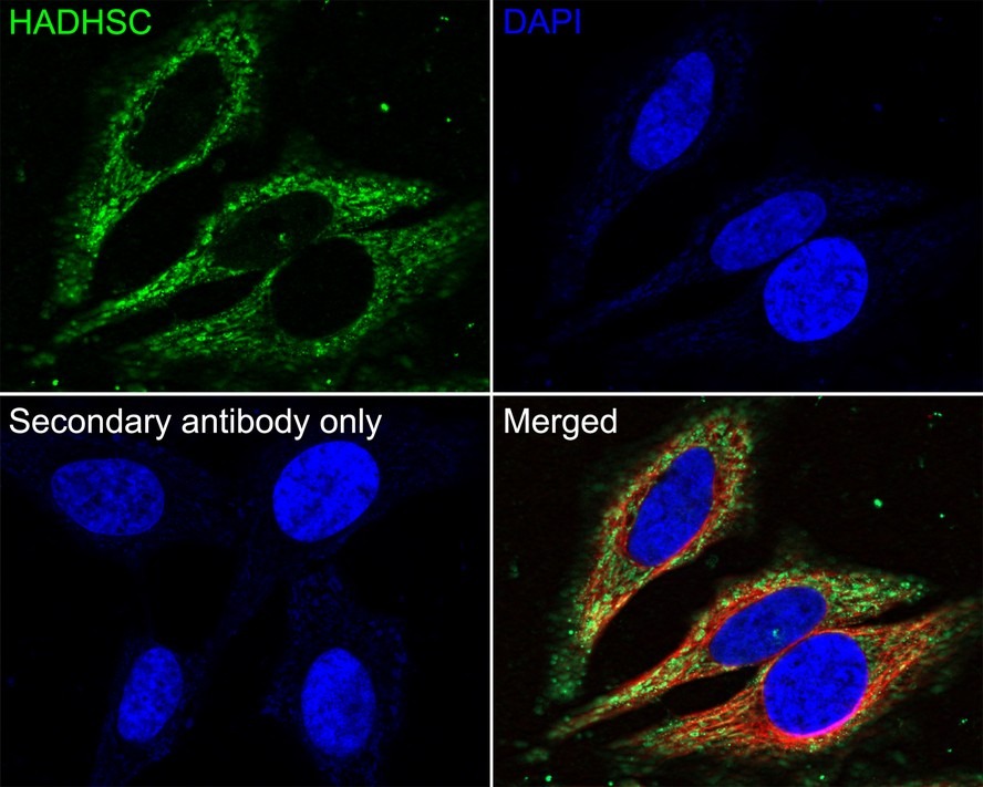 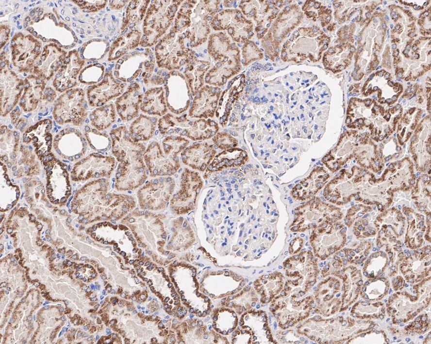 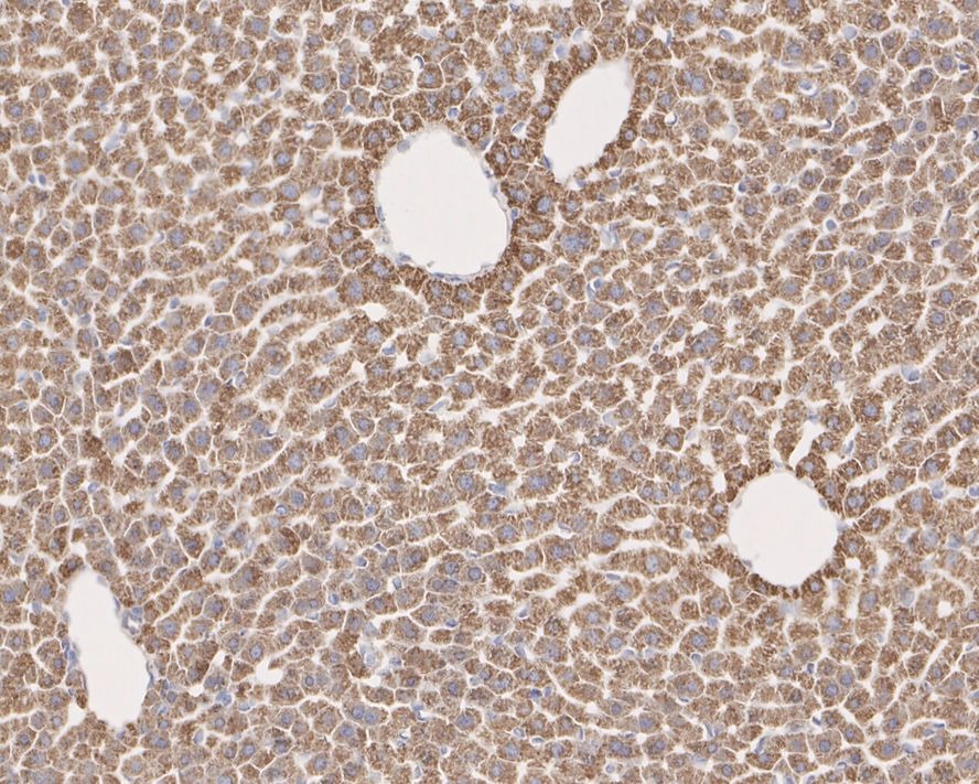 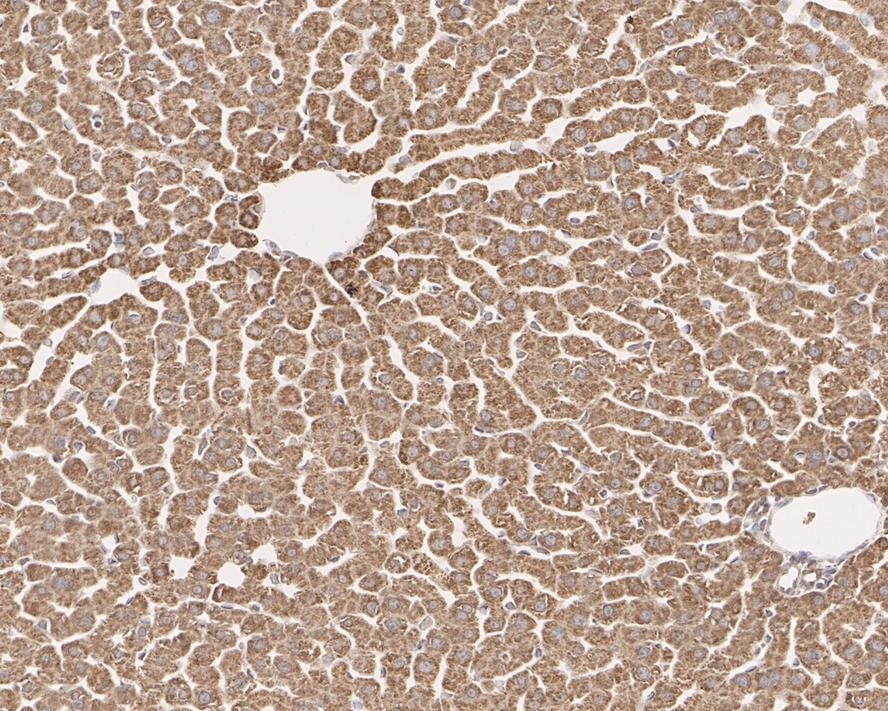 |
| 应用 | ICCIHCWB |
| 推荐剂量 | WB: 1:1000-2000; ICC: 1:500 |
| 抗体种类 | Monoclonal |
| 宿主来源 | Mouse |
| 构建方式 | Hybridoma Monoclonal Antibody |
| 纯化方式 | ProA affinity purified |
| 性状 | Liquid |
| 缓冲液 | 1*TBS (pH7.4), 0.5%BSA, 40%Glycerol. Preservative: 0.05% Sodium Azide. |
| 研究背景 | Hydroxyacyl-Coenzyme A dehydrogenase also known as HADH is an enzyme which in humans is encoded by the HADH gene. This gene is a member of the 3-hydroxyacyl-CoA dehydrogenase gene family. The encoded protein functions in the mitochondrial matrix to catalyze the oxidation of straight-chain 3-hydroxyacyl-CoAs as part of the beta-oxidation pathway. Its enzymatic activity is highest with medium-chain-length fatty acids. Mutations in this gene cause one form of familial hyperinsulinemic hypoglycemia. A deficiency is associated with 3-hydroxyacyl-coenzyme A dehydrogenase deficiency. |
| 偶联 | Unconjugated |
| 免疫原 | Peptide |
| Uniprot ID |
| 分子量 | Theoretical: 34 kDa. |
| 储存方式 | Store at -20°C or -80°C for 12 months. Avoid repeated freeze-thaw cycles. |
| 运输方式 | Shipping with blue ice. |
 嗨!有任何问题?点我咨询
版权所有©2015-2026 TargetMol Chemicals Inc.保留所有权利.
沪ICP备20019793号-4 | 沪公网安备 31010602006700号 | 沪(静)应急管危经许[2024]203441